Call us
301-363-4651 (Available 9 a.m. to 5 p.m. CST from Monday to Friday)
| Code | CSB-RA002611A0HU |
| Size | US$210 |
| Order now | |
| Image |
|
| Have Questions? | Leave a Message or Start an on-line Chat |
| Application | Recommended Dilution |
|---|---|
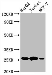
| WB | 1:500-1:5000 |
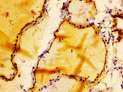
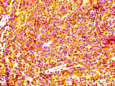
| IHC | 1:50-1:500 |
BCL2 stands as one of the most extensively studied regulators of programmed cell death, functioning as a critical anti-apoptotic protein that prevents mitochondrial outer membrane permeabilization. Its overexpression is implicated in numerous malignancies, where it enables cancer cells to evade apoptotic signals, making it an essential target for researchers investigating cell survival mechanisms, therapeutic resistance, and oncogenic pathways.
This recombinant monoclonal antibody, generated from clone 2C6 in rabbit host, offers the reproducibility that apoptosis research demands. Because the antibody sequence is defined and production occurs through recombinant expression, you can expect consistent performance across experiments and between lot numbers, eliminating a common source of variability when tracking subtle changes in BCL2 expression levels.
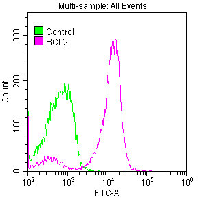
Validation data demonstrates reliable detection across multiple experimental platforms. Western blot analysis confirms specific recognition of BCL2 at the expected 26 kDa molecular weight in HepG2 hepatocellular carcinoma, Jurkat T-cell leukemia, and MCF-7 breast adenocarcinoma cell lysates, providing confidence when working with diverse cancer models. Immunohistochemical staining has been optimized in paraffin-embedded human thyroid and lymph node tissues using citrate buffer antigen retrieval, enabling examination of BCL2 expression patterns in tissue architecture. Flow cytometry validation in Jurkat cells further extends utility to single-cell analysis of intracellular BCL2 levels following fixation and permeabilization.
With cross-species reactivity encompassing human, mouse, and rat samples, this antibody supports translational studies moving between model systems and human specimens. Whether you are characterizing apoptotic resistance in tumor samples, evaluating therapeutic responses, or investigating developmental cell death programs, this antibody provides a dependable tool for BCL2 detection across your experimental workflow.
Applications : Immunohistochemical staining
Sample type: tissue
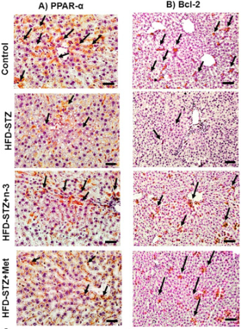
Review: Effects of n3-PUFAs (n-3, 300 mg/kg) and metformin (Met, 150 mg/kg) on relative hepatic tissue expression of peroxisome proliferator-activated receptor alpha (PPAR-α) and B-cell lymphoma 2 (Bcl-2) in high-fat diet/low dose streptozotocin (HFD-STZ)-induced NAFLD in rats by immunohistochemistry. (×400, scale bar = 50um).
By Anonymous

