
Call us
301-363-4651 (Available 9 a.m. to 5 p.m. CST from Monday to Friday)

Cadherins are a family of Ca²⁺-dependent transmembrane cell adhesion molecules essential for tissue organization in multicellular organisms. Beyond their canonical role in cell–cell adhesion, cadherins participate in tissue development, cell polarity establishment, mechanotransduction, signal transduction, and organ homeostasis.
Recent studies have highlighted their involvement in tumor invasion, angiogenesis, autoimmune diseases, developmental disorders, and tissue fibrosis. Given their tissue-specific expression patterns and functional diversity, cadherin family members have emerged as promising targets for disease mechanism research, biomarker development, and therapeutic intervention. This review systematically summarizes the structural features, functional classifications, and biological mechanisms of the cadherin superfamily, with a focus on representative members in tissue homeostasis and disease pathogenesis. We also discuss current research tools and translational applications to inform future basic research and drug discovery efforts.
Cadherins were originally discovered as cell–cell adhesion molecules that maintain multicellular tissue integrity. Through homophilic binding between extracellular domains on adjacent cells, cadherins form stable adhesive junctions that preserve the spatial organization of epithelial, endothelial, cardiac, and neural tissues. For this reason, cadherins have long been regarded as the "molecular glue" of tissue stability.
However, cadherins do far more than physically tether cells together. Their intracellular domains interact with p120-catenin, β-catenin, and α-catenin to form a protein complex that links adhesion to the cytoskeleton and multiple signaling pathways (Fig. 1). This enables cells to sense environmental changes and dynamically regulate proliferation, migration, differentiation, and apoptosis. Thus, cadherins function not only as structural anchors but also as critical platforms for coordinating cellular behavior.
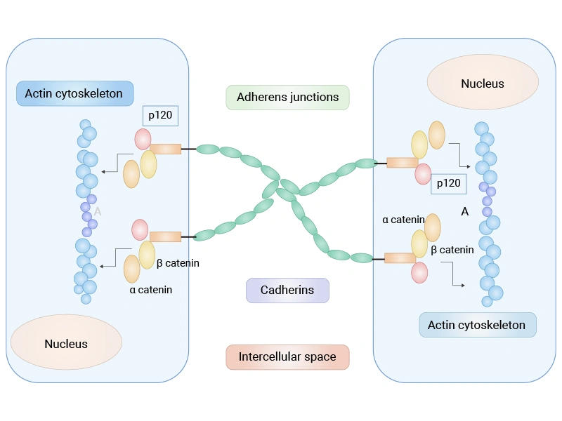
Figure 1. Schematic of the cadherin–catenin complex at adherens junctions [23]
This structure–signaling coupling places cadherins at the center of tissue homeostasis. When cell–cell junctions are intact, cadherins maintain tissue polarity, restrict aberrant migration, and suppress sustained pro-proliferative signaling. When cadherin expression is reduced, mutated, or mislocalized, adhesion weakens and tissue architecture destabilizes, while multiple signaling pathways become reactivated—ultimately driving tumor invasion, fibrosis, and developmental abnormalities.
During evolution, the cadherin superfamily has diversified into distinct functional branches. Classical cadherins maintain epithelial, endothelial, and neural connections; desmosomal cadherins provide mechanical reinforcement in skin and myocardium; and non-classical cadherins primarily regulate neural circuit formation, planar cell polarity, and organ development. Despite their functional differences, all family members rely on cadherin-mediated adhesion as a common foundation for tissue homeostasis and disease regulation.
Understanding cadherin biology requires not only appreciating their adhesive functions but also recognizing how they integrate cell junctions, mechanical stimuli, and signal transduction into a unified regulatory network.
Cadherin-mediated adhesion is strictly Ca²⁺-dependent. The extracellular region consists of multiple cadherin repeats, with Ca²⁺-binding sites located between adjacent domains. Ca²⁺ binding maintains the extended, rigid conformation required for accurate homophilic trans-binding between neighboring cells. When local Ca²⁺ concentration drops, the extracellular domains collapse and adhesive activity is lost [1].
This reversible mechanism allows cadherins to both stabilize tissues and dynamically remodel cell–cell connections in response to injury, inflammation, and development.
Cadherin function depends not only on extracellular adhesion but also on the intracellular protein complex. Through p120-catenin, β-catenin, and α-catenin, cadherins link to the actin cytoskeleton, forming a continuous mechanical network that enables both structural coupling and force transmission between cells.
Recent studies have revealed that this complex acts as a mechanosensor. Upon mechanical stimuli such as stretch, compression, or shear stress, junctional proteins undergo conformational changes and recruit additional cytoskeletal components, reinforcing intercellular connections. This adaptive response is essential for the long-term stability of skin, blood vessels, and cardiac tissue under continuous mechanical load [2].
Beyond adhesion, cadherins regulate multiple signaling pathways. The Wnt/β-catenin pathway is the best-characterized example. Under normal conditions, most β-catenin is sequestered at the membrane by the cadherin complex, limiting its nuclear availability. When cadherin expression declines or junctions are disrupted, β-catenin is released, translocates to the nucleus, and activates TCF/LEF-dependent transcription, driving proliferation, migration, and phenotypic remodeling [3,4].
Cadherins also crosstalk with Hippo-YAP, Rho GTPase, and PI3K/Akt signaling networks. By integrating adhesion status, mechanical cues, and microenvironmental signals, they coordinate cell polarity, proliferation, differentiation, and migration. Thus, cadherins serve as both structural scaffolds and signaling platforms that link mechanical forces to cell fate decisions—a common molecular foundation for the tissue-specific functions of different family members [3].
Classical cadherins are the most extensively studied subgroup, represented by CDH1 (E-cadherin), CDH2 (N-cadherin), and CDH5 (VE-cadherin). These molecules are characterized by their involvement in tissue architecture and cell phenotype transitions, forming a regulatory axis between epithelial and mesenchymal states.
CDH1 is predominantly expressed in epithelial tissues and is the core component of adherens junctions. By linking adjacent cells to the actin cytoskeleton, CDH1 maintains cell polarity, barrier integrity, and tissue organization.
CDH1 also functions as a negative regulator of Wnt/β-catenin signaling. Under normal conditions, CDH1 anchors β-catenin to the membrane, restricting its nuclear entry. When CDH1 expression is reduced, mutated, or silenced by promoter methylation, β-catenin is released, translocates to the nucleus, and activates oncogenes such as c-Myc and Cyclin D1, while simultaneously relieving constraints on cell migration. This makes CDH1 loss a key initiating event in epithelial–mesenchymal transition (EMT) [3,4].
Clinically, CDH1 germline mutations are a major cause of hereditary diffuse gastric cancer, and reduced expression is frequently observed in breast, gastric, and liver cancers [5]. Current research on CDH1 focuses on restoring its expression, inhibiting its degradation, and utilizing soluble E-cadherin (sE-cadherin) as a disease monitoring biomarker [6].
CDH2 is primarily expressed in mesenchymal cells, neural tissues, and myocardium, where it plays important roles in tissue remodeling, synaptic formation, and cell–cell interactions. Compared to CDH1, CDH2-mediated junctions are more dynamic, facilitating cell migration and tissue reorganization.
During tumor progression, the downregulation of CDH1 and upregulation of CDH2—known as the "cadherin switch"—is a hallmark of EMT. This switch weakens stable epithelial junctions, enhances migration, invasion, and metastatic capacity, and cooperates with TGF-β and FGF signaling to promote tumor progression [7].
Given its direct role in tumor invasion, CDH2 has become a promising therapeutic target. ADH-1 (Exherin), a cyclic peptide antagonist targeting the CDH2 extracellular domain, is a representative drug candidate, and several function-blocking antibodies are in preclinical development for cancer and fibrotic diseases [8].
CDH5 is almost exclusively expressed in vascular endothelial cells and is essential for maintaining vascular barrier integrity. Under normal conditions, CDH5 forms continuous endothelial junctions that restrict plasma leakage and inflammatory cell extravasation.
Upon VEGF stimulation, the CDH5 intracellular domain undergoes phosphorylation, weakening its interaction with catenins and transiently loosening endothelial junctions to increase vascular permeability. This is a normal physiological response during angiogenesis but becomes pathological when persistently activated in tumors, inflammation, and sepsis, leading to vascular leakage, edema, and abnormal vessel formation [9].
Current research on CDH5 focuses on regulating vascular permeability, improving drug delivery, and inhibiting pathological angiogenesis. Circulating endothelial cells and soluble VE-cadherin are also being explored as potential disease monitoring biomarkers [9].
Desmosomal cadherins include the Desmoglein (DSG1–4) and Desmocollin (DSC1–3) families. Unlike classical cadherins, which link to actin, desmosomal cadherins connect to intermediate filaments (keratin or desmin) via Plakoglobin, Plakophilin, and Desmoplakin, forming exceptionally strong intercellular junctions. They are therefore abundant in skin and myocardium—tissues that endure continuous mechanical stress.
Given their role in resisting tensile and shear forces, desmosomal abnormalities typically manifest not as increased cell migration but as reduced tissue mechanical stability, leading to blistering skin diseases, cardiomyopathies, and autoimmune disorders.
DSG2 is the most widely expressed desmosomal cadherin in cardiac tissue and is essential for connecting adjacent cardiomyocytes. Together with Desmoplakin and Plakoglobin, DSG2 forms stable desmosomal complexes that link desmin intermediate filaments into a continuous network, allowing mechanical stress to be uniformly transmitted across the myocardium during contraction.
Loss-of-function mutations in DSG2 destabilize desmosomal structures, weaken intercellular coupling, and ultimately lead to cardiomyocyte apoptosis, fibrofatty replacement, and conduction abnormalities—hallmarks of arrhythmogenic cardiomyopathy (ACM), which carries high risks of malignant arrhythmias and sudden death [10].
No DSG2-targeted therapies are currently approved, but AAV-mediated gene supplementation has shown promise in restoring desmosomal structure and improving cardiac function in animal models.
DSG3 is predominantly expressed in the basal layer of skin and oral mucosa, where it maintains robust intercellular adhesion in stratified squamous epithelia.
In pemphigus vulgaris (PV), autoantibodies recognize the DSG3 extracellular domain, directly blocking its homophilic adhesion and rapidly dismantling desmosomal junctions, leading to keratinocyte separation (acantholysis) and blister formation [11]. Because this process is antibody-driven, DSG3 has become a classic model for studying autoimmunity.
Anti-DSG3 IgG detection is now a standard serological diagnostic and disease activity monitoring tool for PV. Furthermore, chimeric autoantibody receptor T cells (CAAR-T) engineered with the DSG3 extracellular domain can selectively eliminate pathogenic B cells, representing one of the most promising precision therapeutic strategies for PV, currently in early clinical translation [12].
Beyond classical and desmosomal cadherins, the superfamily includes a large and structurally diverse group of non-classical members, primarily protocadherins (PCDHs), the FAT family, and the CELSR family. Most of these do not directly participate in typical cell–cell adhesion but instead regulate neural circuit formation, planar cell polarity, organ development, and tissue morphogenesis—making them major research foci in developmental biology and genetics.
The protocadherin family is the largest branch of the cadherin superfamily, comprising clustered PCDHs (PCDHA, PCDHB, PCDHG) and non-clustered PCDHs. Unlike classical cadherins, PCDHs lack typical catenin-binding sites in their intracellular domains and are primarily involved in neuronal recognition and connectivity rather than stable mechanical adhesion.
Clustered PCDHs are expressed in a stochastic combinatorial manner, endowing each neuron with a unique surface identity that enables self/non-self discrimination and prevents inappropriate connections—a mechanism essential for proper neural circuit assembly [13].
Among PCDH family members, PCDH19 and PCDH15 are the most studied. PCDH19 mutations cause clustering epilepsy (CE), with disease mechanisms involving PCDH19–β-catenin interaction and Wnt pathway dysregulation [14]. PCDH15 mutations cause Usher syndrome type 1F, a leading cause of hereditary deaf–blindness, for which AAV-mediated gene therapy is now in preclinical development [20].
The FAT family comprises FAT1–FAT4, characterized by exceptionally large extracellular domains that mediate long-range cell interactions. Recent studies have shown that FAT proteins not only participate in cell adhesion but also regulate Hippo-YAP signaling and planar cell polarity, playing important roles in organ development, tissue morphogenesis, and tumorigenesis.
FAT1 is highly expressed in glioblastoma and other malignancies, where it promotes tumor progression by suppressing autophagic cell death [15]. FAT1 also exhibits high-frequency mutations in head and neck squamous cell carcinoma, correlating with immunotherapy resistance [21]. Additionally, FAT1 inactivation promotes YAP/TAZ stabilization by relieving degradation [22].
FAT4 functions as a tumor suppressor and is involved in neuronal migration and organ development. Germline FAT4 mutations cause Van Maldergem syndrome, underscoring the essential role of FAT family members in embryonic development.
CELSR1–CELSR3 are seven-pass transmembrane proteins that serve as receptors in the planar cell polarity (PCP) pathway. Unlike classical cadherins that primarily maintain cell junctions, CELSR family members govern directional cell alignment and coordinated movement within the tissue plane, with critical roles in neural tube closure, neuronal migration, ciliary orientation, and cardiovascular development.
CELSR1 mutations have been linked to brain malformations, neurodevelopmental disorders, and epilepsy [16]. With advances in single-cell sequencing and spatial transcriptomics, the functional networks of CELSR family members in neural development and organogenesis continue to be refined, positioning this family as a major focus of future developmental biology research.
Several cadherin family members exhibit restricted tissue expression patterns. Their high expression in specific organs or diseases, combined with favorable target selectivity, has made them increasingly attractive for therapeutic development in oncology, fibrosis, and tissue repair.
CDH17, also known as LI-cadherin, is primarily expressed in small intestinal and colonic epithelium, where it maintains digestive epithelial integrity and participates in substance transport. Unlike classical cadherins, CDH17 has a short intracellular domain and mediates adhesion independently of the catenin complex, giving it unique biological properties.
CDH17 is consistently overexpressed in colorectal, gastric, and some pancreatic cancers, correlating with invasive capacity and patient prognosis [17]. Its relatively restricted expression in normal tissues makes it an attractive therapeutic target. ADC and bispecific antibody-based therapies targeting CDH17 are showing promise in colorectal and esophageal adenocarcinoma, with ongoing clinical studies.
CDH11 is primarily expressed in fibroblasts, mesenchymal cells, and bone tissue, hence its alternative name OB-cadherin. Recent studies have shown that CDH11 not only participates in bone formation but also plays broad roles in post-inflammatory tissue remodeling and fibrosis.
CDH11 expression is significantly elevated in pulmonary fibrosis, rheumatoid arthritis, and osteoarthritis, where it promotes fibroblast activation, extracellular matrix deposition, and chronic inflammation. As such, CDH11 has emerged as a promising anti-fibrotic target, with several monoclonal antibodies showing efficacy in animal models [18].
CDH6 is involved in kidney and embryonic development, with limited expression in adult normal tissues but sustained high expression in ovarian, renal clear cell, and some thyroid cancers, making it an attractive tumor-selective target.
With the rapid advancement of ADC technology, CDH6 has become a next-generation ADC target. HKT288, a CDH6-targeting ADC, has demonstrated potent antitumor activity in ovarian cancer models and is now in clinical translation [19]. Its low expression in normal tissues offers advantages in therapeutic window expansion and off-target toxicity reduction, positioning CDH6 as a notable target for precision therapy in gynecological cancers.
Beyond these, additional tissue-specific cadherins are emerging as novel targets for disease research and drug development, with their translational potential expected to expand further.
Recent advances have revealed the expanding roles of the cadherin family in oncology, biomechanics, developmental biology, and immunology. An increasing number of cadherin members are not only serving as tools for understanding disease mechanisms but also evolving into diagnostic biomarkers and therapeutic targets.
To support cadherin-related research, CUSABIO offers a range of high-quality recombinant proteins, antibodies, and ELISA kits covering multiple family members. These products are suitable for protein expression analysis, functional mechanism studies, and biomarker detection, providing experimental support for both basic research and drug development.
| Target | Code | Product Name | Source |
|---|---|---|---|
| CDH1 | CSB-EP005034HU | Recombinant Human Cadherin-1 (CDH1), partial | E.coli |
| CDH1 | CSB-MP005034HU1 | Recombinant Human Cadherin-1 (CDH1), partial (Active) | Mammalian cell |
| CDH1 | CSB-MP5601MOV | Recombinant Macaca fascicularis Cadherin-1 (CDH1), partial (Active) | Mammalian cell |
| CDH10 | CSB-MP897318HU | Recombinant Human Cadherin-10 (CDH10), partial | Mammalian cell |
| CDH12 | CSB-EP005037HUc7 | Recombinant Human Cadherin-12 (CDH12), partial | E.coli |
| CDH12 12 | CSB-EP005037HU | Recombinant Human Cadherin-12 (CDH12), partial | E.coli |
| CDH16 | CSB-BP005041HU | Recombinant Human Cadherin-16 (CDH16), partial | Baculovirus |
| Cdh16 | CSB-EP005041MO | Recombinant Mouse Cadherin-16 (Cdh16), partial | E.coli |
| Cdh16 | CSB-MP005041MO | Recombinant Mouse Cadherin-16 (Cdh16), partial | Mammalian cell |
| Cdh17 | CSB-EP882608MO | Recombinant Mouse Cadherin-17 (Cdh17), partial | E.coli |
| Cdh17 | CSB-YP882608MO | Recombinant Mouse Cadherin-17 (Cdh17), partial | Yeast |
| CDH17 | CSB-MP613267HU | Recombinant Human Cadherin-17 (CDH17), partial (Active) | Mammalian cell |
| CDH17 | CSB-MP4664MOV | Recombinant Macaca fascicularis Cadherin 17 (CDH17), partial (Active) | Mammalian cell |
| Cdh17 | CSB-MP882608MO1 | Recombinant Mouse Cadherin-17 (Cdh17), partial | Mammalian cell |
| CDH17 | CSB-MP613267HUh8 | Recombinant Human Cadherin-17 (CDH17), partial | Mammalian cell |
| Cdh17 | CSB-MP005042RA | Recombinant Rat Cadherin-17 (Cdh17), partial (Active) | Mammalian cell |
| CDH18 | CSB-MP615709HU | Recombinant Human Cadherin-18 (CDH18), partial | Mammalian cell |
| CDH19 | CSB-MP887951HU | Recombinant Human Cadherin-19 (CDH19), partial | Mammalian cell |
| CDH20 | CSB-MP864022HU | Recombinant Human Cadherin-20 (CDH20), partial | Mammalian cell |
| CDH3 | CSB-YP005052PI | Recombinant Pig Cadherin-3 (CDH3) | Yeast |
| Target | Code | Product Name | Tested Applications |
|---|---|---|---|
| CDH1 | CSB-RA576116A0HU | CDH1 Recombinant Monoclonal Antibody | ELISA, IHC |
| CDH1 | CSB-RA005034MA1HU | CDH1 Recombinant Monoclonal Antibody | ELISA, WB, IHC, FC |
| CDH1 | CSB-MA196808 | CDH1 Monoclonal Antibody | ELISA, IHC |
| CDH1 | CSB-MA005034A0m | CDH1 Monoclonal Antibody | ELISA, WB, IF, FC |
| CDH1 | CSB-PA080147 | CDH1 Antibody | WB, IHC |
| CDH1 | CSB-PA002243 | CDH1 Antibody | WB, IHC, IF, ELISA |
| CDH1 | CSB-PA005034GA01HU | CDH1 Antibody | ELISA, WB, IF |
| CDH1 | CSB-PA218378 | CDH1 Antibody | ELISA, WB, IHC |
| CDH1 | CSB-PA164023 | CDH1 Antibody | ELISA, WB, IHC |
| CDH1 | CSB-PA908796 | CDH1 Antibody | ELISA, IHC |
| CDH1 | CSB-PA938817 | CDH1 Antibody | ELISA, IHC |
| CDH1 | CSB-PA795479 | CDH1 Antibody | ELISA, WB |
| CDH1 | CSB-PA06239A0Rb | CDH1 Antibody | ELISA, IHC, IF |
| CDH1 | CSB-PA06239B0Rb | CDH1 Antibody, HRP conjugated | ELISA |
| CDH1 | CSB-PA06239C0Rb | CDH1 Antibody, FITC conjugated | N/A |
| CDH1 | CSB-PA06239D0Rb | CDH1 Antibody, Biotin conjugated | ELISA |
| CDH1 | CSB-PA005034MA01HU | CDH1 Antibody | ELISA, WB, IHC, IF, IP |
| CDH1 | CSB-PA005034MB01HU | CDH1 Antibody, HRP conjugated | ELISA |
| CDH1 | CSB-PA005034MC01HU | CDH1 Antibody, FITC conjugated | N/A |
| CDH1 | CSB-PA005034MD01HU | CDH1 Antibody, Biotin conjugated | ELISA |
| Target | Code | Product Name | Detection Range | Sensitivity |
|---|---|---|---|---|
| CDH1 | CSB-E04519h | Human E-Cadherin,E-Cad ELISA Kit | 0.312 ng/ml -20 ng/ml | 0.078 ng/ml |
| CDH1 | CSB-E04520m | Mouse E-Cadherin,E-Cad ELISA Kit | 0.312 ng/ml-20 ng/ml | 0.078ng/ml |
| CDH1 | CSB-E07308r | Rat Epithelial-Cadherin,E-Cad ELISA Kit | 3.12 ng/mL-200 ng/mL | 0.78 ng/mL |
| CDH13 | CSB-E13817h | Human H-Cadherin,H-Cad/CDH13 ELISA Kit | 1.25 ng/mL-80 ng/mL | 0.312 ng/mL |
| CDH2 | CSB-E09718h | Human Neural -Cadherin, N-Cad ELISA kit | 6.25 ng/mL-400 ng/mL | 1.56 ng/mL |
| CDH3 | CSB-E08955h | Human Placenta Cadherin,P-cad ELISA Kit | / | / |
| CDH3 | CSB-EL005052MO | Mouse Cadherin-3(CDH3) ELISA kit | 0.625 ng/ml-40 ng/ml | 0.156ng/ml |
| CDH5 | CSB-E09372h | Human Vascular Endothelial-Cadherin,VE-cad ELISA Kit | 3.9 ng/mL-250 ng/mL | 0.98 ng/mL |
| CDH5 | CSB-EL005054MO | Mouse Cadherin-5(CDH5) ELISA kit | 0.235 ng/mL-15 ng/mL | 0.058 ng/mL |
| DSG1 | CSB-E09592h | Human desmogleins 1,Dsg-1 ELISA Kit | 23.5 pg/mL-1500 pg/mL | 5.8 pg/mL |
| DSG3 | CSB-EL007205HU | Human Desmoglein-3(DSG3) ELISA kit | 28 pg/mL-1800 pg/mL | 7 pg/mL |
| PCDH10 | CSB-EL017525HU | Human Protocadherin-10(PCDH10) ELISA kit | 0.312 ng/mL-20 ng/mL | 0.078 ng/mL |
[1] Nagar B,Overduin M,Ikura M, et al. Structural basis of calcium-induced E-cadherin rigidification and dimerization. Nature. 1996;380 (6572):360-4.
[2] Mertz AF,Che Y,Banerjee S, et al. Cadherin-based intercellular adhesions organize epithelial cell-matrix traction forces. Proc Natl Acad Sci U S A. 2013;110 (3):842-7.
[3] Tian X,Liu Z,Niu B, et al. E-cadherin/β-catenin complex and the epithelial barrier. J Biomed Biotechnol. 2011;2011:567305.
[4] Wang N, Wang Y, Zhang L, et al. Molecular Mechanisms of Epithelial–Mesenchymal Transition in Retinal Pigment Epithelial Cells: Implications for Age-Related Macular Degeneration (AMD) Progression. Biomolecules. 2025; 15, 771.
[5] Blair VR,McLeod M,Carneiro F, et al. Hereditary diffuse gastric cancer: updated clinical practice guidelines. Lancet Oncol. 2020;21 (8):e386-e397.
[6] Corso G,Figueiredo J,De Angelis SP, et al. E-cadherin deregulation in breast cancer. J Cell Mol Med. 2020;24 (11):5930-5936.
[7] Loh CY,Chai JY,Tang TF, et al. The E-Cadherin and N-Cadherin Switch in Epithelial-to-Mesenchymal Transition: Signaling, Therapeutic Implications, and Challenges. Cells. 2019;8 (10):.
[8] Khorsand M,Khajeh S,Eslami M, et al. Telmisartan anti-cancer activities mechanism through targeting N-cadherin by mimicking ADH-1 function. J Cell Mol Med. 2022;26 (8):2392-2403.
[9] Nan W,He Y,Wang S, et al. Molecular mechanism of VE-cadherin in regulating endothelial cell behaviour during angiogenesis. Front Physiol. 2023;14:1234104.[10] Debus JD,Milting H,Brodehl A, et al. In vitro analysis of arrhythmogenic cardiomyopathy associated desmoglein-2 (DSG2) mutations reveals diverse glycosylation patterns. J Mol Cell Cardiol. 2019;129:303-313.
[11] Tedbirt B,Calbo S,Lebourgeois L, et al. Persistence of anti-desmoglein 3 antibodies in patients with pemphigus vulgaris in complete remission is associated with restricted isotype diversity and desmoglein 3 ectodomain recognition. Br J Dermatol. 2026;194 (5):959-961.
[12] Ellebrecht CT,Bhoj VG,Nace A, et al. Reengineering chimeric antigen receptor T cells for targeted therapy of autoimmune disease. Science. 2016;353 (6295):179-84.
[13] Takeichi M. The cadherin superfamily in neuronal connections and interactions. Nat Rev Neurosci. 2007;8 (1):11-20.
[14] de Nys R,de Nys R,Gardner A, et al. Proteomic analysis of the developing mammalian brain links PCDH19 to the Wnt/β-catenin signalling pathway. Mol Psychiatry. 2024;29 (7):2199-2210.
[15] Gupta Y,Goswami S,Arora M, et al. Atypical cadherin FAT1 promotes tumorigenesis by suppressing autophagic cell death in glioblastoma under hypoxia or nutrient stress. Cell Mol Biol Lett. 2025;30 (1):106.
[16] Bonardi CM,Ruiz-Reig N,Chai G, et al. Biallelic variants in CELSR1 cause brain malformations, neurodevelopmental disorders and epilepsy in humans. Nat Commun. 2026;17 (1):862.
[17] Pushpadevan R,Viehweger F,Gorbokon N, et al. Cadherin-17 (CDH17) expression in human cancer: A tissue microarray study on 18,131 tumors. Pathol Res Pract. 2024;256:155175.
[18] Pedroza M , George T , Agarwal S .Cadherin-11 regulates pulmonary fibrosis in bleomycin-induced lung injury (MUC5P.865)[J].The Journal of Immunology, 2014, 192(1_Supplement):134.8-134.8.
[19] Saxena P,Dürr C,Prieur P, et al. Discovery and Optimization of HKT288, a Cadherin-6-Targeting ADC for the Treatment of Ovarian and Renal Cancers. Cancer Discov. 2017;7 (9):1030-1045.
[20] Hathaway DM,Klein AJ,Pan B, et al. Mini-PCDH15 gene therapy rescues hearing in a mouse model of Usher syndrome type 1F. Nat Commun. 2023;14 (1):2400.
[21] Cao H,Lan T,Kuang S, et al. FAT1 as a tumor mutation burden specific gene affects the immunotherapy effect in head and neck squamous cell cancer. Drug Resist Updat. 2024;76:101095.
[22] Li R,Shao J,Jin YJ, et al. Endothelial FAT1 inhibits angiogenesis by controlling YAP/TAZ protein degradation via E3 ligase MIB2. Nat Commun. 2023;14 (1):1980.
[23] Sisto M,Ribatti D,Lisi S. Cadherin Signaling in Cancer and Autoimmune Diseases. Int J Mol Sci. 2021;22 (24):.


