Call us
301-363-4651 (Available 9 a.m. to 5 p.m. CST from Monday to Friday)
Argonaute 2 (AGO2), also known as eukaryotic translation initiation factor 2C2 (EIF2C2), is the catalytic core component of the RNA-induced silencing complex (RISC). It mediates post-transcriptional gene silencing via cleaving target mRNAs or repressing translation, a central function in the RNA interference (RNAi) pathway that regulates cell proliferation, differentiation, and apoptosis.
Dysregulated AGO2 expression is linked to cancers (e.g., breast, lung) where it modulates oncogenic miRNA-mRNA networks to promote tumorigenesis. Altered AGO2 activity also disrupts miRNA homeostasis in neurodegenerative diseases like Alzheimer’s. Current drug development targets its endonuclease activity or optimizes RNAi-based therapies dependent on its function, with preclinical progress but limited clinical trials to date.
Recombinant Human Protein argonaute-2 (AGO2), partial (CSB-EP891731HU)
Validated Data
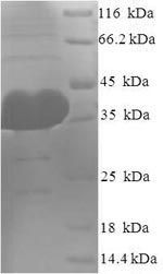
(Tris-Glycine gel) Discontinuous SDS-PAGE (reduced) with 5% enrichment gel and 15% separation gel.
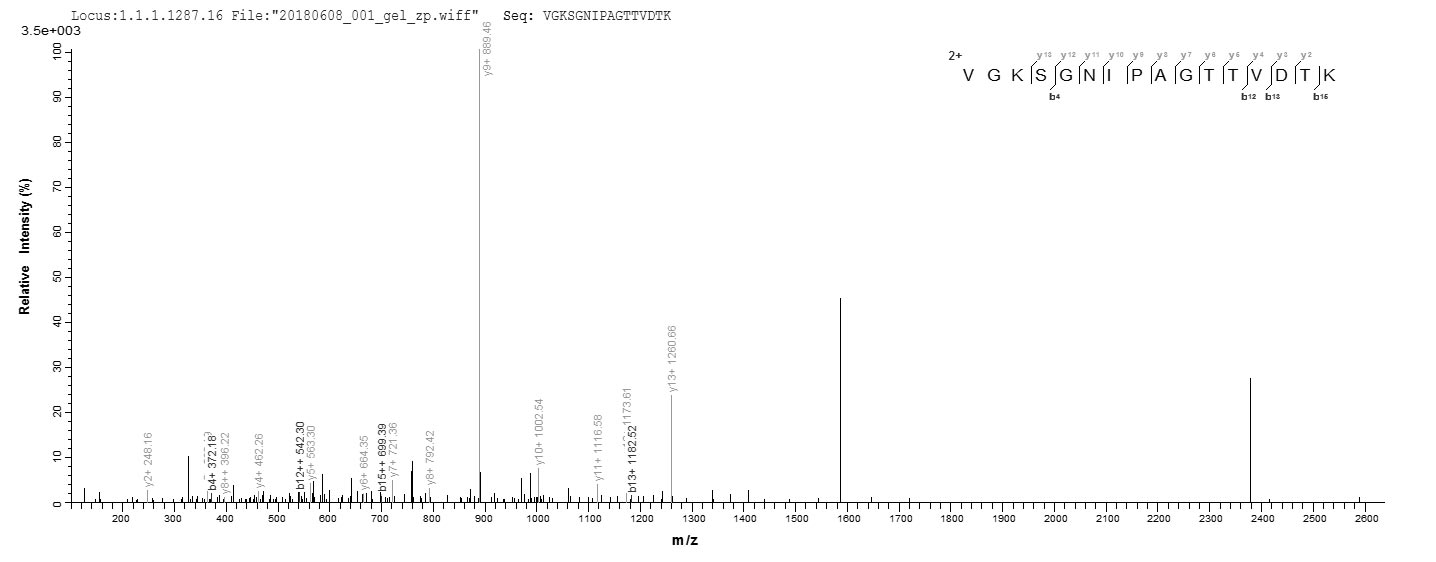
Based on the SEQUEST from database of E.coli host and target protein, the LC-MS/MS Analysis result of CSB-EP891731HU could indicate that this peptide derived from E.coli-expressed Homo sapiens (Human) AGO2.
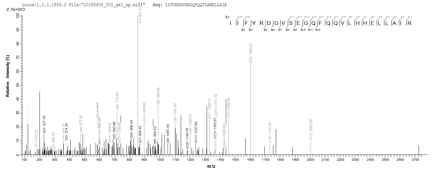
Based on the SEQUEST from database of E.coli host and target protein, the LC-MS/MS Analysis result of CSB-EP891731HU could indicate that this peptide derived from E.coli-expressed Homo sapiens (Human) AGO2.
AGO2 Recombinant Monoclonal Antibody (CSB-RA965823A0HU)
Validated Data
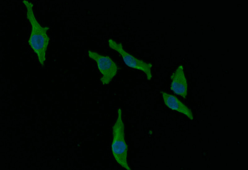
Immunofluorescence staining of MCF-7 with CSB-RA965823A0HU at 1:30, counter-stained with DAPI. The cells were fixed in 4% formaldehyde and blocked in 10% normal Goat Serum. The cells were then incubated with the antibody overnight at 4°C. The secondary antibody was Alexa Fluor 492-congugated AffiniPure Goat Anti-Rabbit IgG(H+L).
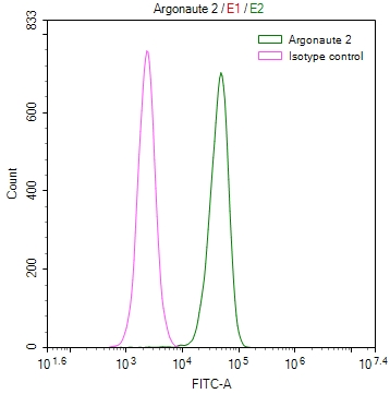
Overlay Peak curve showing Hela cells stained with CSB-RA965823A0HU (red line) at 1:50. The cells were fixed in 4% formaldehyde and permeated by 0.2% TritonX-100. Then 10% normal goat serum to block non-specific protein-protein interactions followed by the antibody (1µg/1*106cells) for 45min at 4℃. The secondary antibody used was FITC-conjugated Goat Anti-rabbit IgG(H+L) at 1:200 dilution for 35min at 4℃.Control antibody (green line) was rabbit IgG (1µg/1*106cells) used under the same conditions. Acquisition of >10,000 events was performed.
AGO2 Antibody (CSB-PA891731LA01HU)
Validated Data
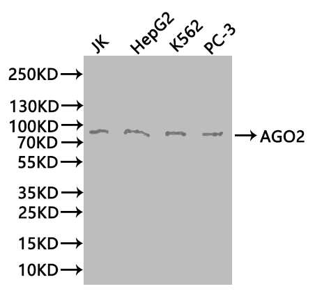
Western Blot
Positive WB detected in: JK whole cell lysate(20µg), HepG2 whole cell lysate(20µg), K562 whole cell lysate(20µg), PC-3 whole cell lysate(20µg)
All lanes: AGO2 antibody at 1:1000
Secondary
Goat polyclonal to rabbit IgG at 1/50000 dilution
Predicted band size: 98 kDa
Observed band size: 98 kDa
Exposure time:1min
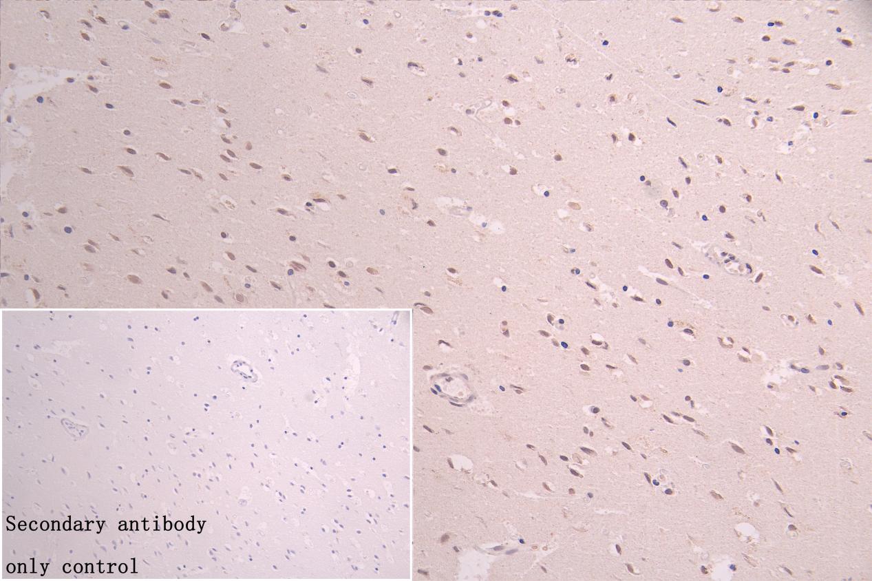
IHC image of CSB-PA891731LA01HU diluted at 1:800 and staining in paraffin-embedded human brain tissue performed on a Leica BondTM system. After dewaxing and hydration, antigen retrieval was mediated by high pressure in a citrate buffer (pH 6.0). Section was blocked with 10% normal goat serum 30min at RT. Then primary antibody (1% BSA) was incubated at 4°C overnight. The primary is detected by a Goat anti-rabbit polymer IgG labeled by HRP and visualized using 0.05% DAB. Secondary antibody only control: uses 1% BSA instead of primary antibody
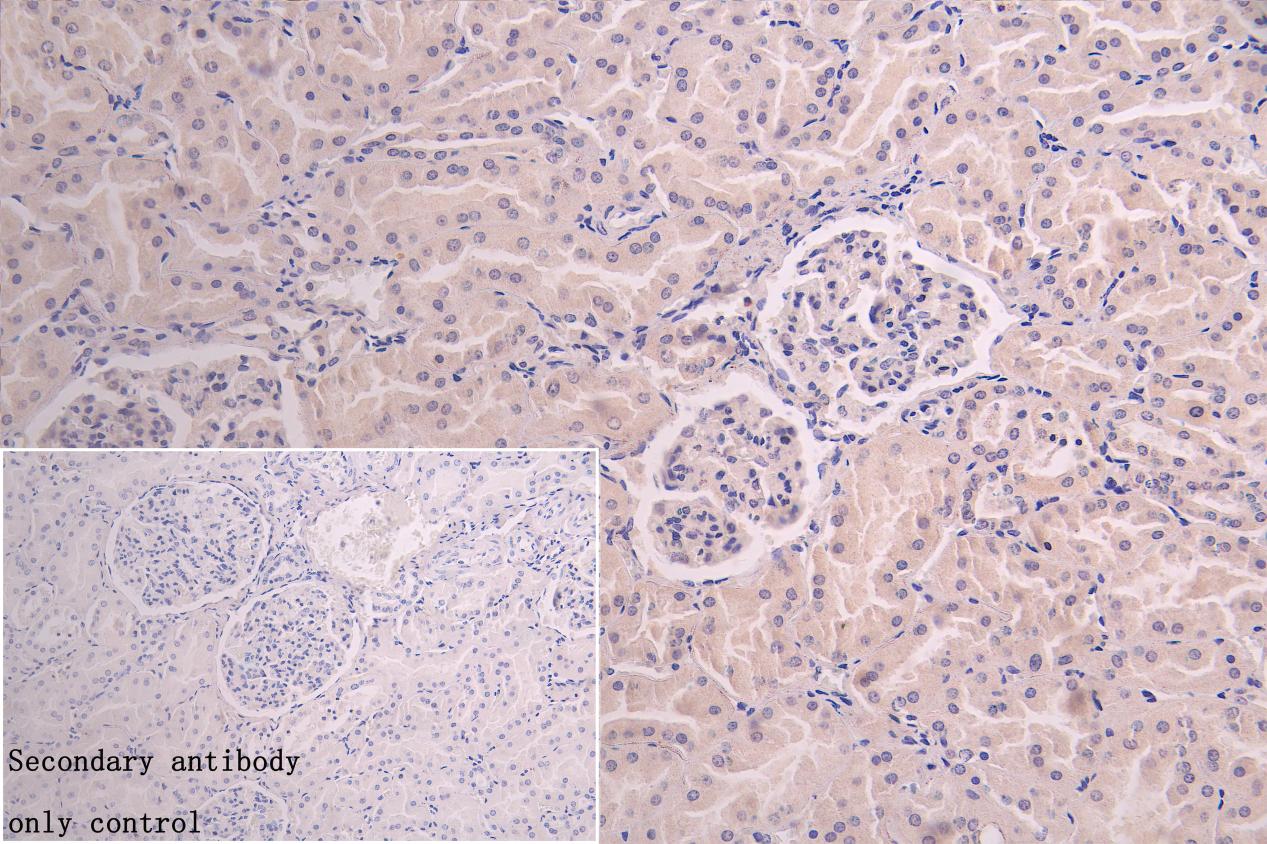
IHC image of CSB-PA891731LA01HU diluted at 1:800 and staining in paraffin-embedded human kidney tissue performed on a Leica BondTM system. After dewaxing and hydration, antigen retrieval was mediated by high pressure in a citrate buffer (pH 6.0). Section was blocked with 10% normal goat serum 30min at RT. Then primary antibody (1% BSA) was incubated at 4°C overnight. The primary is detected by a Goat anti-rabbit polymer IgG labeled by HRP and visualized using 0.05% DAB. Secondary antibody only control: uses 1% BSA instead of primary antibody
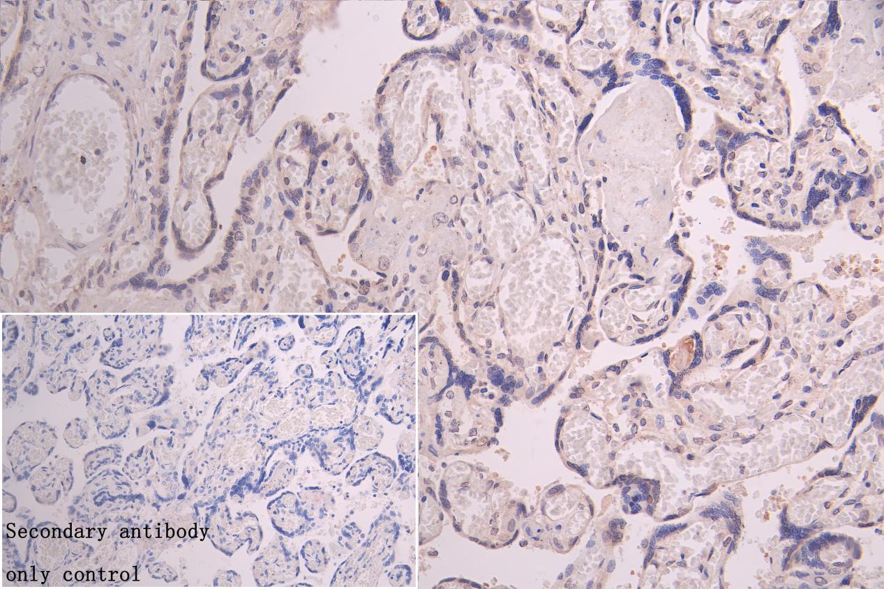
IHC image of CSB-PA891731LA01HU diluted at 1:800 and staining in paraffin-embedded human placenta tissue performed on a Leica BondTM system. After dewaxing and hydration, antigen retrieval was mediated by high pressure in a citrate buffer (pH 6.0). Section was blocked with 10% normal goat serum 30min at RT. Then primary antibody (1% BSA) was incubated at 4°C overnight. The primary is detected by a Goat anti-rabbit polymer IgG labeled by HRP and visualized using 0.05% DAB. Secondary antibody only control: uses 1% BSA instead of primary antibody

Chromatin Immunoprecipitation Hela (1.1*106) were cross-linked with formaldehyde, sonicated,and immunoprecipitated with 4µg anti-AgO2 or a control normal rabbit IgG. The resulting ChIP DNA was quantified tissue using real-time PCR with primers (CSB-PP891731HU) against the SLC1A5 promoter.
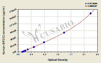
Human Protein argonaute-2(EIF2C2) ELISA kit (CSB-EL007520HU)
Validated Data
Code: CSB-EL007520HU
Size: 96T,5×96T,10×96T
Sensitivity: 6.25 pg/mL
Detection Range: 25 pg/mL-1600 pg/mL
These standard curves are provided for demonstration only. A standard curve should be generated for each set of samples assayed.
The following AGO2 reagents supplied by CUSABIO are manufactured under a strict quality control system. Multiple applications have been validated and solid technical support is offered.
AGO2 Antibodies for Homo sapiens (Human)
| Code | Product Name | Species Reactivity | Application |
|---|---|---|---|
| CSB-PA891731LC01HU | AGO2 Antibody, FITC conjugated | Human | |
| CSB-PA891731LD01HU | AGO2 Antibody, Biotin conjugated | Human | ELISA |
| CSB-PA290707 | AGO2 Antibody | Human,Mouse,Rat | ELISA,IHC |
| CSB-PA891731LA01HU | AGO2 Antibody | Human, Mouse | ELISA, WB, IHC, ChIP |
| CSB-RA965823A0HU | AGO2 Recombinant Monoclonal Antibody | Human | ELISA, IF, FC |
AGO2 Proteins for Homo sapiens (Human)
| Code | Product Name | Source |
|---|---|---|
| CSB-EP891731HU | Recombinant Human Protein argonaute-2 (AGO2), partial | E.coli |
| CSB-YP891731HU | Recombinant Human Protein argonaute-2 (AGO2), partial | Yeast |
| CSB-BP891731HU CSB-MP891731HU CSB-EP891731HU-B |
Recombinant Human Protein argonaute-2 (AGO2), partial | Baculovirus Mammalian cell In Vivo Biotinylation in E.coli |
AGO2 ELISA Kit for Homo sapiens (Human)
| Code | Product Name | Sample Types | Sensitivity |
|---|---|---|---|
| CSB-EL007520HU | Human Protein argonaute-2(EIF2C2) ELISA kit | serum, plasma, tissue homogenates, cell lysates | 6.25 pg/mL |

