Call us
301-363-4651 (Available 9 a.m. to 5 p.m. CST from Monday to Friday)
CDK6, cell cycle protein-dependent kinase 6, alias cytokinin 6, platelet-derived serine/threonine kinase, and others. It is a protein kinase encoded by the CDK6 gene, a member of the CMGC family of protein kinases, which is important for the progression of the G1 phase of the cell cycle and the G1/S transition.CDK6 binds to D-type cell cycle proteins to form a complex that promotes the entry of cells into the S phase for DNA replication by phosphorylating the retinoblastoma protein (Rb) and activating the E2F transcriptional program.
CDK6 The biological significance of CDK6 is mainly reflected in its role in the regulation of the cell cycle. Its activity is essential for maintaining normal cell proliferation, while CDK6 is also involved in cell differentiation and tumor cell proliferation. In tumor cells, aberrant activation of CDK6 has been associated with the development of a variety of cancers, including leukemia, lymphoma, and solid tumors. In addition, CDK6 inhibitors such as Palbociclib, Ribociclib, and Abemaciclib have been used clinically for the treatment of certain types of breast cancer and are being investigated for the treatment of other cancer types.
CDK6 Recombinant Monoclonal Antibody (CSB-RA555745A0HU)
Validated Data
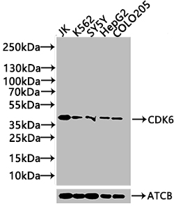
Western Blot
Positive WB detected in: JK whole cell lysate(20µg), K562 whole cell lysate(20µg), SY5Y whole cell lysate(20µg), HepG2 whole cell lysate(20µg), COLO205 whole cell lysate(20µg)
All lanes: CDK6 antibody at 1:1000
Secondary
Goat polyclonal to rabbit IgG at 1/40000 dilution
Predicted band size: 37 kDa
Observed band size: 37 kDa
Exposure time: 120s
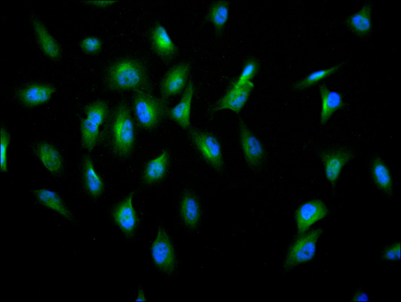
Immunofluorescence staining of U251 cell with CSB-RA555745A0HU at 1:10, counter-stained with DAPI. The cells were fixed in 4% formaldehyde and and permeated by 0.2% TritonX-100 for 15 min. Then 10% normal goat serum to block non-specific protein-protein interactions . The cells were then incubated with the antibody overnight at 4℃. The secondary antibody was Alexa Fluor 488-congugated AffiniPure Goat Anti-Rabbit IgG(H+L).
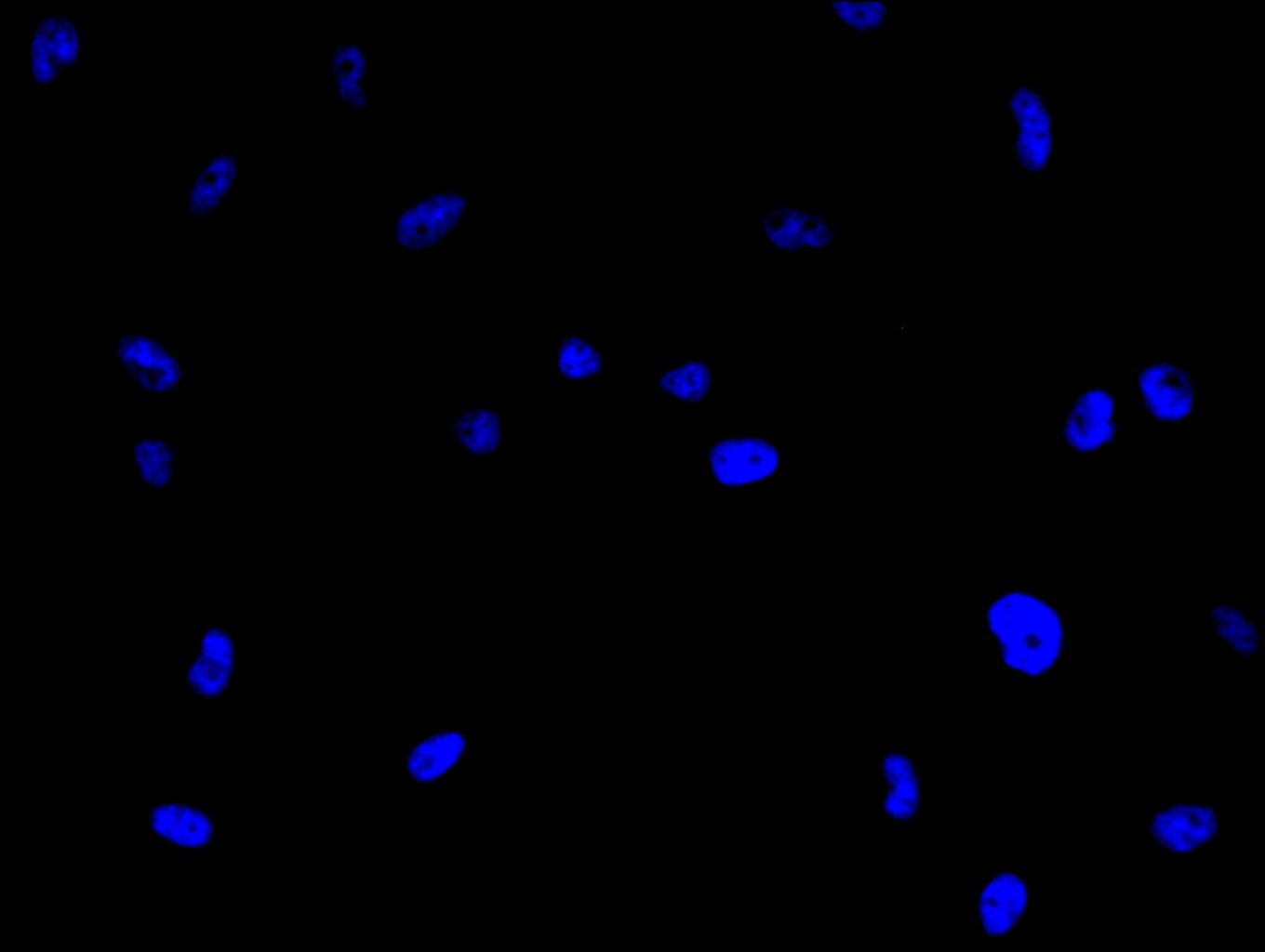
Immunofluorescence staining of U251 cell with 5% goat serum, counter-stained with DAPI. The cells were fixed in 4% formaldehyde and blocked in 10% normal Goat Serum. The cells were then incubated with the antibody overnight at 4C. The secondary antibody was Alexa Fluor 488-congugated AffiniPure Goat Anti-Rabbit IgG(H+L).
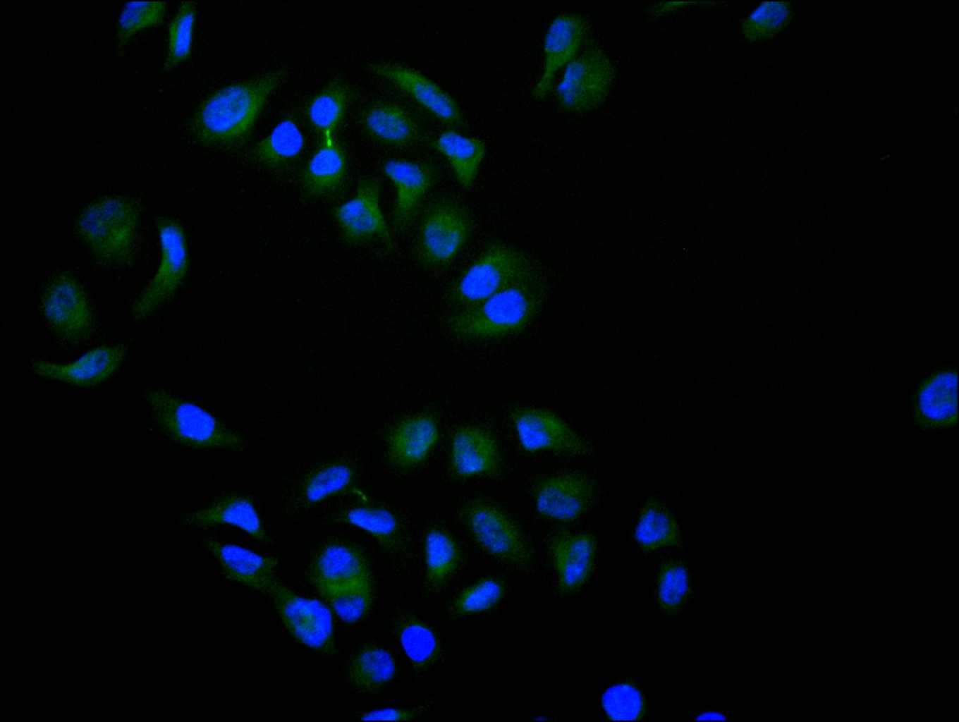
Immunofluorescence staining of Hela cell with CSB-RA555745A0HU at 1:10, counter-stained with DAPI. The cells were fixed in 4% formaldehyde and and permeated by 0.2% TritonX-100 for 15 min. Then 10% normal goat serum to block non-specific protein-protein interactions . The cells were then incubated with the antibody overnight at 4℃. The secondary antibody was Alexa Fluor 488-congugated AffiniPure Goat Anti-Rabbit IgG(H+L).
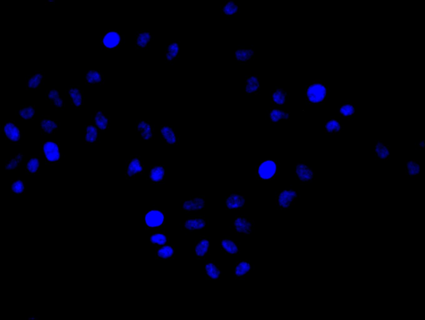
Immunofluorescence staining of Hela cell with 5% goat serum, counter-stained with DAPI. The cells were fixed in 4% formaldehyde and blocked in 10% normal Goat Serum. The cells were then incubated with the antibody overnight at 4C. The secondary antibody was Alexa Fluor 488-congugated AffiniPure Goat Anti-Rabbit IgG(H+L).
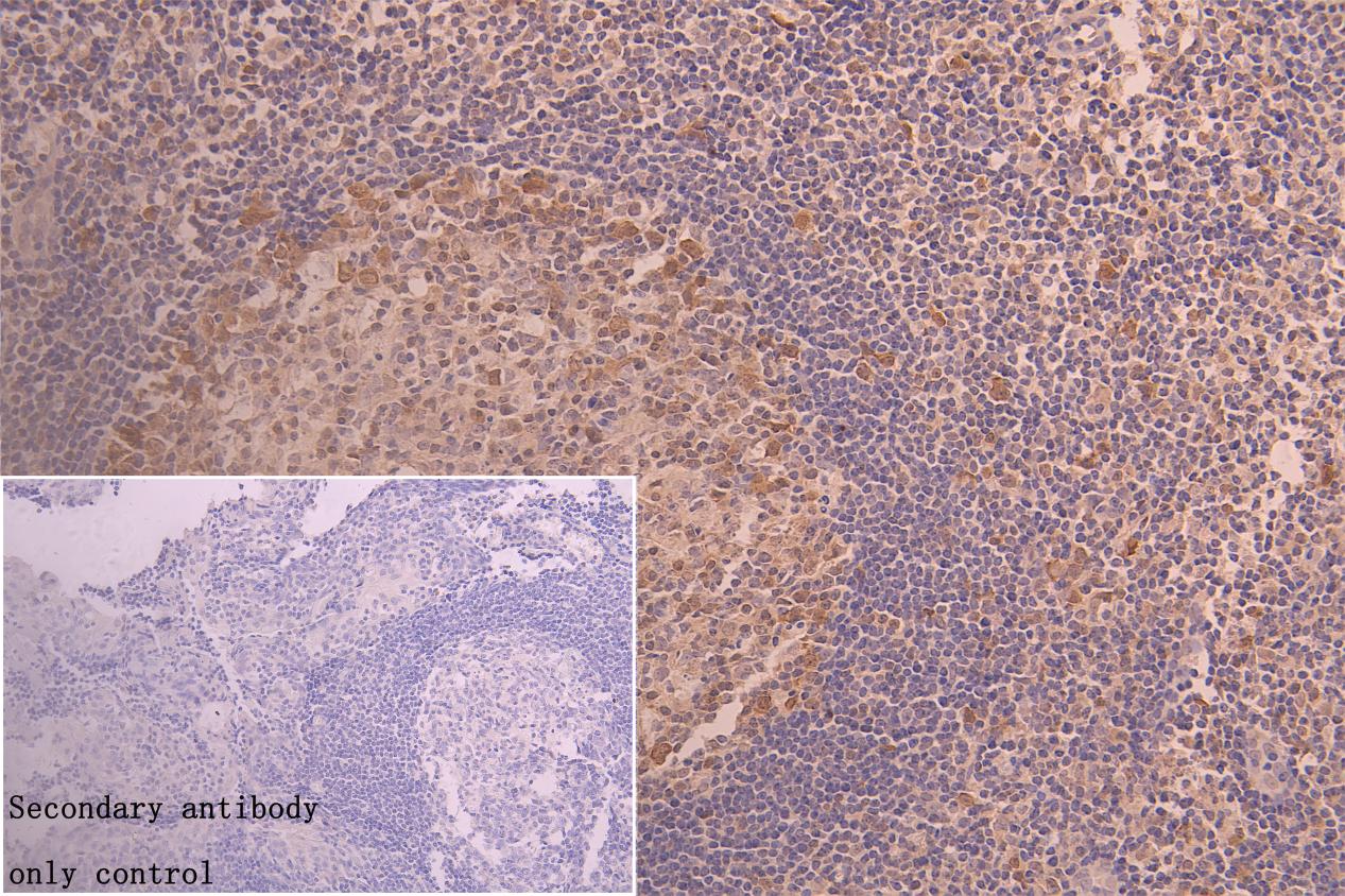
IHC image of CSB-RA555745A0HU diluted at 1:50 and staining in paraffin-embedded human tonsil tissue performed on a Leica BondTM system. After dewaxing and hydration, antigen retrieval was mediated by high pressure in a citrate buffer (pH 6.0). Section was blocked with 10% normal goat serum 30min at RT. Then primary antibody (1% BSA) was incubated at 4°C overnight. The primary is detected by a Goat anti-rabbit polymer IgG labeled by HRP and visualized using 0.05% DAB. Secondary antibody only control: uses 1% BSA instead of primary antibody
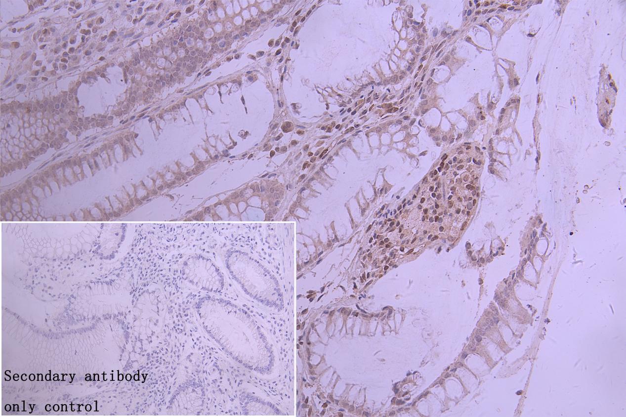
IHC image of CSB-RA555745A0HU diluted at 1:50 and staining in paraffin-embedded human colorectal cancer performed on a Leica BondTM system. After dewaxing and hydration, antigen retrieval was mediated by high pressure in a citrate buffer (pH 6.0). Section was blocked with 10% normal goat serum 30min at RT. Then primary antibody (1% BSA) was incubated at 4°C overnight. The primary is detected by a Goat anti-rabbit polymer IgG labeled by HRP and visualized using 0.05% DAB. Secondary antibody only control: uses 1% BSA instead of primary antibody
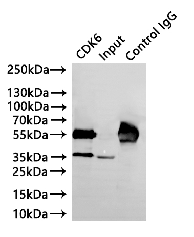
Immunoprecipitating CDK6 in K562whole cell lysate
Lane 1: CSB-RA555745A0HU(3µg)+ K562 whole cell lysate(220µg)
Lane 2: K562 whole cell lysate(30µg)
Lane 3:Rabbit control IgG instead of CSB-RA555745A0HU in K562 whole cell lysate
For western blotting, a HRP-conjugated Protein G antibody was used as the secondary antibody (1/40000)
The following CDK6 reagents supplied by CUSABIO are manufactured under a strict quality control system. Multiple applications have been validated and solid technical support is offered.
CDK6 Antibodies for Homo sapiens (Human)
| Code | Product Name | Species Reactivity | Application |
|---|---|---|---|
| CSB-PA005074GA01HU | CDK6 Antibody | Human,Mouse,Rat | ELISA,WB |
| CSB-PA549404 | Phospho-CDK6 (Tyr13) Antibody | Human,Mouse | ELISA,WB,IHC,IF |
| CSB-PA267478 | Phospho-CDK6 (Tyr24) Antibody | Human,Mouse | ELISA,WB,IHC,IF |
| CSB-PA090625 | CDK6 (Ab-13) Antibody | Human,Mouse | ELISA,IHC,IF |
| CSB-PA564539 | CDK6 (Ab-24) Antibody | Human,Mouse | ELISA,WB,IHC |
| CSB-PA871419 | CDK6 Antibody | Human,Mouse | ELISA,WB,IHC |
| CSB-PA245607 | CDK6 Antibody | Human | WB, IHC, ELISA |
| CSB-PA005074LA01HU | CDK6 Antibody | Human | ELISA, IHC, IF |
| CSB-PA005074LB01HU | CDK6 Antibody, HRP conjugated | Human | ELISA |
| CSB-PA005074LC01HU | CDK6 Antibody, FITC conjugated | Human | |
| CSB-PA005074LD01HU | CDK6 Antibody, Biotin conjugated | Human | ELISA |
| CSB-RA555745A0HU | CDK6 Recombinant Monoclonal Antibody | Human | ELISA, WB, IHC, IF, IP |
CDK6 Proteins for Mus musculus (Mouse)
| Code | Product Name | Source |
|---|---|---|
| CSB-YP005074MO CSB-EP005074MO CSB-BP005074MO CSB-MP005074MO CSB-EP005074MO-B |
Recombinant Mouse Cyclin-dependent kinase 6 (Cdk6) | Yeast E.coli Baculovirus Mammalian cell In Vivo Biotinylation in E.coli |

