Call us
301-363-4651 (Available 9 a.m. to 5 p.m. CST from Monday to Friday)
PAM, full name peptidylglycine alpha-amidating monooxygenase, is commonly known as PAL, PHM or PAM-1. It is a bifunctional transmembrane enzyme widely expressed in multiple tissues, catalyzing the conversion of inactive peptidylglycine precursors into bioactive alpha-amidated peptides, and participating in copper homeostasis and neuroendocrine regulation.
Abnormal PAM expression or function is associated with type 2 diabetes, cervical cancer, hepatic cirrhosis and pituitary tumors. Currently, research focuses on its role in peptide modification and disease pathogenesis, while targeted modulators and related therapeutic strategies are under preclinical exploration to intervene in related diseases.
PAM Antibody (CSB-PA017417LA01HU)
Validated Data
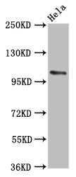
Western Blot
Positive WB detected in: Hela whole cell lysate
All lanes: PAM antibody at 5.6µg/ml
Secondary
Goat polyclonal to rabbit IgG at 1/50000 dilution
Predicted band size: 109, 97, 101, 99, 107 kDa
Observed band size: 109 kDa
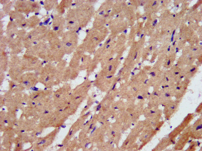
IHC image of CSB-PA017417LA01HU diluted at 1:200 and staining in paraffin-embedded human heart tissue performed on a Leica BondTM system. After dewaxing and hydration, antigen retrieval was mediated by high pressure in a citrate buffer (pH 6.0). Section was blocked with 10% normal goat serum 30min at RT. Then primary antibody (1% BSA) was incubated at 4°C overnight. The primary is detected by a biotinylated secondary antibody and visualized using an HRP conjugated SP system.
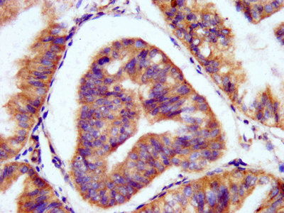
IHC image of CSB-PA017417LA01HU diluted at 1:200 and staining in paraffin-embedded human endometrial cancer performed on a Leica BondTM system. After dewaxing and hydration, antigen retrieval was mediated by high pressure in a citrate buffer (pH 6.0). Section was blocked with 10% normal goat serum 30min at RT. Then primary antibody (1% BSA) was incubated at 4°C overnight. The primary is detected by a biotinylated secondary antibody and visualized using an HRP conjugated SP system.
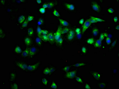
Immunofluorescence staining of Hela cells with CSB-PA017417LA01HU at 1:66, counter-stained with DAPI. The cells were fixed in 4% formaldehyde, permeabilized using 0.2% Triton X-100 and blocked in 10% normal Goat Serum. The cells were then incubated with the antibody overnight at 4°C. The secondary antibody was Alexa Fluor 488-congugated AffiniPure Goat Anti-Rabbit IgG(H+L).
The following PAM reagents supplied by CUSABIO are manufactured under a strict quality control system. Multiple applications have been validated and solid technical support is offered.
PAM Antibodies for Homo sapiens (Human)
| Code | Product Name | Species Reactivity | Application |
|---|---|---|---|
| CSB-PA017417LA01HU | PAM Antibody | Human | ELISA, WB, IHC, IF |
| CSB-PA017417LB01HU | PAM Antibody, HRP conjugated | Human | ELISA |
| CSB-PA017417LC01HU | PAM Antibody, FITC conjugated | Human | |
| CSB-PA017417LD01HU | PAM Antibody, Biotin conjugated | Human | ELISA |
PAM Proteins for Homo sapiens (Human)
| Code | Product Name | Source |
|---|---|---|
| CSB-YP017417HU CSB-EP017417HU CSB-BP017417HU CSB-MP017417HU CSB-EP017417HU-B |
Recombinant Human Peptidyl-glycine alpha-amidating monooxygenase (PAM), partial | Yeast E.coli Baculovirus Mammalian cell In Vivo Biotinylation in E.coli |

