Call us
301-363-4651 (Available 9 a.m. to 5 p.m. CST from Monday to Friday)
Prosaposin, encoded by the PSAP gene, is the precursor of four saposins (A-D) and a secreted neurotrophic factor. Its aliases include SAP precursor and sulfated glycoprotein 1. Saposins assist lysosomal enzymes in sphingolipid degradation, while full-length prosaposin promotes neuronal survival via PI3K/Akt and MAPK pathways.
PSAP mutations cause sphingolipid storage disorders like Gaucher-like phenotypes; its dysregulation links to Alzheimer’s and Parkinson’s. Research focuses on enzyme replacement for storage disorders and neuroprotective agents targeting its trophic activity, but no approved drugs are available yet.
Recombinant Human Proactivator polypeptide (PSAP), partial (CSB-EP018836HU)
Validated Data
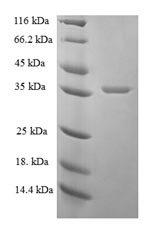
(Tris-Glycine gel) Discontinuous SDS-PAGE (reduced) with 5% enrichment gel and 15% separation gel.
PSAP Antibody (CSB-PA018836DA01HU)
Validated Data
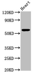
Western Blot
Positive WB detected in: Mouse heart tissue
All lanes: PSAP antibody at 3.3µg/ml
Secondary
Goat polyclonal to rabbit IgG at 1/50000 dilution
Predicted band size: 59 kDa
Observed band size: 59 kDa
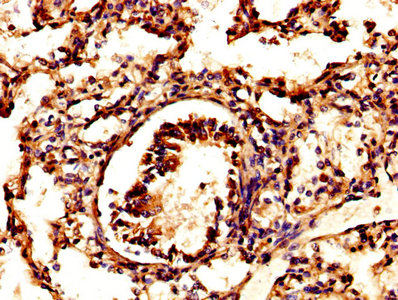
IHC image of CSB-PA018836DA01HU diluted at 1:500 and staining in paraffin-embedded human lung tissue performed on a Leica BondTM system. After dewaxing and hydration, antigen retrieval was mediated by high pressure in a citrate buffer (pH 6.0). Section was blocked with 10% normal goat serum 30min at RT. Then primary antibody (1% BSA) was incubated at 4°C overnight. The primary is detected by a biotinylated secondary antibody and visualized using an HRP conjugated SP system.
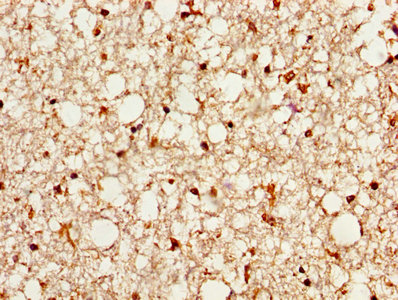
IHC image of CSB-PA018836DA01HU diluted at 1:500 and staining in paraffin-embedded human brain tissue performed on a Leica BondTM system. After dewaxing and hydration, antigen retrieval was mediated by high pressure in a citrate buffer (pH 6.0). Section was blocked with 10% normal goat serum 30min at RT. Then primary antibody (1% BSA) was incubated at 4°C overnight. The primary is detected by a biotinylated secondary antibody and visualized using an HRP conjugated SP system.
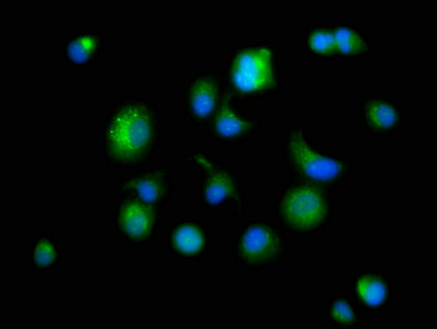
Immunofluorescence staining of MCF-7 cells with CSB-PA018836DA01HU at 1:166, counter-stained with DAPI. The cells were fixed in 4% formaldehyde, permeabilized using 0.2% Triton X-100 and blocked in 10% normal Goat Serum. The cells were then incubated with the antibody overnight at 4°C. The secondary antibody was Alexa Fluor 488-congugated AffiniPure Goat Anti-Rabbit IgG(H+L).
The following PSAP reagents supplied by CUSABIO are manufactured under a strict quality control system. Multiple applications have been validated and solid technical support is offered.
PSAP Antibodies for Homo sapiens (Human)
| Code | Product Name | Species Reactivity | Application |
|---|---|---|---|
| CSB-PA018836DC01HU | PSAP Antibody, FITC conjugated | Human | |
| CSB-PA018836DA01HU | PSAP Antibody | Human, Mouse | ELISA, WB, IHC, IF |
| CSB-PA018836GA01HU | PSAP Antibody | Human,Mouse,Rat | ELISA,WB,IHC |
| CSB-PA040013 | PSAP Antibody | Human | WB, IHC, ELISA |
PSAP Proteins for Homo sapiens (Human)
| Code | Product Name | Source |
|---|---|---|
| CSB-YP018836HU | Recombinant Human Prosaposin (PSAP), partial | Yeast |
| CSB-EP018836HU | Recombinant Human Proactivator polypeptide (PSAP), partial | E.coli |
| CSB-BP018836HU CSB-MP018836HU CSB-EP018836HU-B |
Recombinant Human Proactivator polypeptide (PSAP) | Baculovirus Mammalian cell In Vivo Biotinylation in E.coli |
PSAP ELISA Kit for Homo sapiens (Human)
| Code | Product Name | Sample Types | Sensitivity |
|---|---|---|---|
| CSB-E12837h | human prosaposin(PSAP)Elisa kit | serum, plasma, tissue homogenates. | 0.041 ng/ml |

