Call us
301-363-4651 (Available 9 a.m. to 5 p.m. CST from Monday to Friday)
The protein encoded by TUFM is mitochondrial Tu translation elongation factor, also commonly referred to as EF-Tumt. It plays a crucial role in mitochondrial protein synthesis by facilitating the delivery of aminoacyl-tRNA to the ribosomal A site during translation elongation, utilizing GTP hydrolysis for energy.
TUFM is involved in the mitochondrial translation pathway and interacts with mitochondrial ribosomal subunits. Dysregulation of TUFM has been linked to mitochondrial diseases, including Leigh syndrome and mitochondrial encephalomyopathy. Currently, there are no specific drugs targeting TUFM, but research focuses on understanding its role in mitochondrial dysfunction for potential therapeutic development.
Recombinant Human Elongation factor Tu, mitochondrial (TUFM) (CSB-EP025342HUa0)
Validated Data
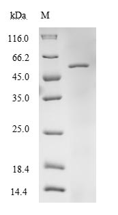
(Tris-Glycine gel) Discontinuous SDS-PAGE (reduced) with 5% enrichment gel and 15% separation gel.
TUFM Recombinant Monoclonal Antibody (CSB-RA025342MA1HU)
Validated Data
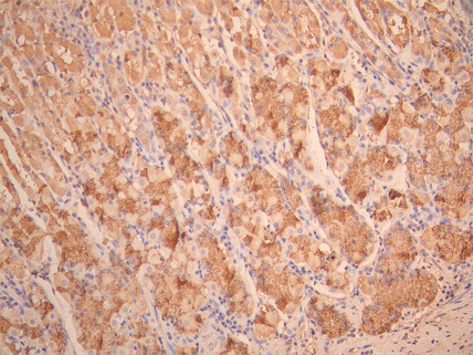
IHC image of CSB-RA025342MA1HU diluted at 1:50 and staining in paraffin-embedded human stomach tissue performed on a Leica BondTM system. After dewaxing and hydration, antigen retrieval was mediated by high pressure in a citrate buffer (pH 6.0). Section was blocked with 10% normal goat serum 30min at RT. Then primary antibody (1% BSA) was incubated at 4°C overnight. The primary is detected by a Anti-Human lgG, Fcy Fragment Specific labeled by HRP and visualized using 0.05% DAB.
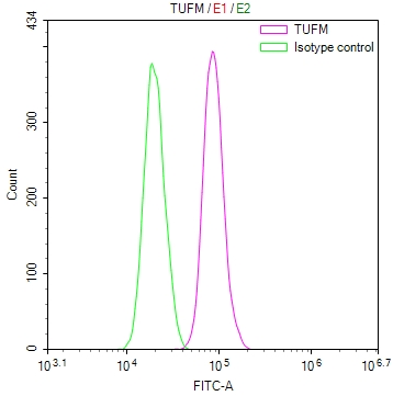
Overlay Peak curve showing Hela cells stained with CSB-RA025342MA1HU (red line) at 1:100. The cells were fixed in 4% formaldehyde and permeated by 0.2% TritonX-100 for 10min. Then 10% normal goat serum to block non-specific protein-protein interactions followed by the antibody (1ug/1*106cells) for 45min at 4℃. The secondary antibody used was Fluorescein (FITC) AffiniPure Goat Anti-Human IgG, Fcγ fragment specific at 1:200 dilution for 35min at 4℃.Control antibody (green line) was human IgG1 (1ug/1*106cells) used under the same conditions. Acquisition of >10,000 events was performed.
Validated Data
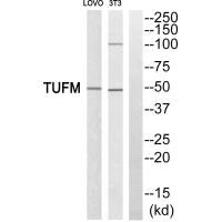
Western blot analysis of extracts from LOVO cells and 3T3 cells, using TUFM antiobdy.
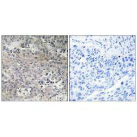
Immunohistochemistry analysis of paraffin-embedded human lung carcinoma tissue using TUFM antibody.
The following TUFM reagents supplied by CUSABIO are manufactured under a strict quality control system. Multiple applications have been validated and solid technical support is offered.
TUFM Antibodies for Homo sapiens (Human)
| Code | Product Name | Species Reactivity | Application |
|---|---|---|---|
| CSB-PA02199A0Rb | TUFM Antibody | Human | ELISA, WB |
| CSB-PA559509 | TUFM Antibody | Human,Mouse | ELISA,WB,IHC |
| CSB-PA060027 | TUFM Antibody | Human,Mouse,Rat | WB, IHC, ELISA |
| CSB-RA025342MA1HU | TUFM Recombinant Monoclonal Antibody | Human | ELISA, IHC, FC |
TUFM Proteins for Homo sapiens (Human)
| Code | Product Name | Source |
|---|---|---|
| CSB-YP025342HU | Recombinant Human Elongation factor Tu, mitochondrial (TUFM) | Yeast |
| CSB-EP025342HU | Recombinant Human Elongation factor Tu, mitochondrial (TUFM) | E.coli |
| CSB-BP025342HU CSB-MP025342HU CSB-EP025342HU-B |
Recombinant Human Elongation factor Tu, mitochondrial (TUFM) | Baculovirus Mammalian cell In Vivo Biotinylation in E.coli |
| CSB-EP025342HU1 | Recombinant Human Elongation factor Tu, mitochondrial (TUFM), partial | E.coli |
| CSB-EP025342HUa0 | Recombinant Human Elongation factor Tu, mitochondrial (TUFM) | E.coli |

