Call us
301-363-4651 (Available 9 a.m. to 5 p.m. CST from Monday to Friday)
CTSS, or histone protease S, alias CathepsinS, Cats, CatS. CTSS is encoded by the human CTSS gene, which is located on chromosome 1 in the region 1q21.3.
CTSS is a lysosomal cysteine protease belonging to the family of peptidases C1, which is involved in the degradation of antigenic proteins into peptides for presentation to MHC class II molecules. CTSS also possesses elastase activity, which extracellularly remodels components of the extracellular matrix.
CTSS has been implicated in the pathology of a variety of inflammatory and autoimmune diseases and plays an important role in certain lung diseases due to its elastase activity. Aberrant expression and regulation of CTSS has been implicated in the pathogenesis of a variety of diseases, including lung diseases, such as pulmonary fibrosis, in which overexpression of CTSS causes multiple pathological processes, including pulmonary fibrosis, increased CTSS secretion and accelerated extracellular matrix remodeling.
Recombinant Human Cathepsin S (CTSS) (CSB-EP006204HU)
Validated Data
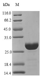
(Tris-Glycine gel) Discontinuous SDS-PAGE (reduced) with 5% enrichment gel and 15% separation gel.
CTSS Recombinant Monoclonal Antibody (CSB-RA788455A0HU)
Validated Data
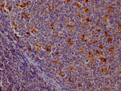
IHC image of CSB-RA788455A0HU diluted at 1:100 and staining in paraffin-embedded human tonsil tissue performed on a Leica BondTM system. After dewaxing and hydration, antigen retrieval was mediated by high pressure in a citrate buffer (pH 6.0). Section was blocked with 10% normal goat serum 30min at RT. Then primary antibody (1% BSA) was incubated at 4℃ overnight. The primary is detected by a Goat anti-rabbit IgG polymer labeled by HRP and visualized using 0.05% DAB.
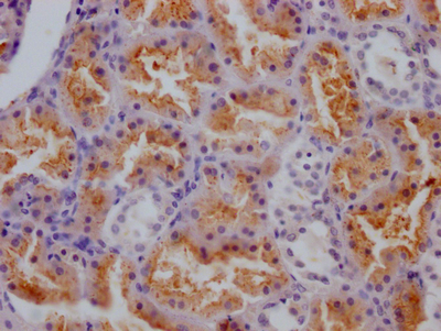
IHC image of CSB-RA788455A0HU diluted at 1:100 and staining in paraffin-embedded human kidney tissue performed on a Leica BondTM system. After dewaxing and hydration, antigen retrieval was mediated by high pressure in a citrate buffer (pH 6.0). Section was blocked with 10% normal goat serum 30min at RT. Then primary antibody (1% BSA) was incubated at 4℃ overnight. The primary is detected by a Goat anti-rabbit IgG polymer labeled by HRP and visualized using 0.05% DAB.
Human Cathepsin S (CTSS) ELISA Kit (CSB-E13722h)
Validated Data
Code: CSB-E13722h
Size: 96T,5×96T,10×96T
Sensitivity: 15.6 pg/mL
Detection Range: 62.5 pg/mL- 4000 pg/mL
These standard curves are provided for demonstration only. A standard curve should be generated for each set of samples assayed.
The following CTSS reagents supplied by CUSABIO are manufactured under a strict quality control system. Multiple applications have been validated and solid technical support is offered.
CTSS Antibodies for Homo sapiens (Human)
| Code | Product Name | Species Reactivity | Application |
|---|---|---|---|
| CSB-PA10729A0Rb | CTSS Antibody | Human | ELISA, WB, IHC |
| CSB-RA788455A0HU | CTSS Recombinant Monoclonal Antibody | Human | ELISA, IHC |
CTSS Antibodies for Mus musculus (Mouse)
| Code | Product Name | Species Reactivity | Application |
|---|---|---|---|
| CSB-PA006204LA01MO | Ctss Antibody | Mouse | ELISA |
| CSB-PA006204LB01MO | Ctss Antibody, HRP conjugated | Mouse | ELISA |
CTSS Proteins for Rattus norvegicus (Rat)
| Code | Product Name | Source |
|---|---|---|
| CSB-YP006204RA CSB-EP006204RA CSB-BP006204RA CSB-MP006204RA CSB-EP006204RA-B |
Recombinant Rat Cathepsin S (Ctss) | Yeast E.coli Baculovirus Mammalian cell In Vivo Biotinylation in E.coli |
CTSS Proteins for Homo sapiens (Human)
| Code | Product Name | Source |
|---|---|---|
| CSB-YP006204HU CSB-BP006204HU CSB-MP006204HU CSB-EP006204HU-B |
Recombinant Human Cathepsin S (CTSS) | Yeast Baculovirus Mammalian cell In Vivo Biotinylation in E.coli |
| CSB-EP006204HU | Recombinant Human Cathepsin S (CTSS) | E.coli |
CTSS Proteins for Bos taurus (Bovine)
| Code | Product Name | Source |
|---|---|---|
| CSB-YP006204BO CSB-EP006204BO CSB-BP006204BO CSB-MP006204BO CSB-EP006204BO-B |
Recombinant Bovine Cathepsin S (CTSS) | Yeast E.coli Baculovirus Mammalian cell In Vivo Biotinylation in E.coli |
CTSS Proteins for Mus musculus (Mouse)
| Code | Product Name | Source |
|---|---|---|
| CSB-YP006204MO CSB-BP006204MO CSB-MP006204MO CSB-EP006204MO-B |
Recombinant Mouse Cathepsin S (Ctss) | Yeast Baculovirus Mammalian cell In Vivo Biotinylation in E.coli |
| CSB-EP006204MO | Recombinant Mouse Cathepsin S (Ctss) | E.coli |
CTSS Proteins for Canis lupus familiaris (Dog) (Canis familiaris)
| Code | Product Name | Source |
|---|---|---|
| CSB-YP006204DO CSB-EP006204DO CSB-BP006204DO CSB-MP006204DO CSB-EP006204DO-B |
Recombinant Dog Cathepsin S (CTSS) | Yeast E.coli Baculovirus Mammalian cell In Vivo Biotinylation in E.coli |
CTSS Proteins for Saimiri boliviensis boliviensis (Bolivian squirrel monkey)
| Code | Product Name | Source |
|---|---|---|
| CSB-YP006204SWC CSB-BP006204SWC CSB-MP006204SWC CSB-EP006204SWC-B |
Recombinant Saimiri boliviensis boliviensis Cathepsin S (CTSS) | Yeast Baculovirus Mammalian cell In Vivo Biotinylation in E.coli |
CTSS ELISA Kit for Homo sapiens (Human)
| Code | Product Name | Sample Types | Sensitivity |
|---|---|---|---|
| CSB-E13722h | Human Cathepsin S (CTSS) ELISA Kit | serum, plasma, tissue homogenates | 15.6 pg/mL |
CTSS ELISA Kit for Mus musculus (Mouse)
| Code | Product Name | Sample Types | Sensitivity |
|---|---|---|---|
| CSB-EL006204MO | Mouse Cathepsin S(CTSS) ELISA kit | serum, plasma, tissue homogenates, cell lysates | 7.8 pg/mL |
CTSS ELISA Kit for Rattus norvegicus (Rat)
| Code | Product Name | Sample Types | Sensitivity |
|---|---|---|---|
| CSB-EL006204RA | Rat Cathepsin S(CTSS) ELISA kit | serum, plasma, tissue homogenates, cell lysates | 3.9 pg/mL |

