Call us
301-363-4651 (Available 9 a.m. to 5 p.m. CST from Monday to Friday)
DLAT (dihydrolipoamide S-acetyltransferase), also known as PDC-E2, is a core subunit of the mitochondrial pyruvate dehydrogenase complex (PDC). It catalyzes acetyl group transfer from dihydrolipoamide to coenzyme A, bridging glycolysis and the tricarboxylic acid cycle, and is critical for mitochondrial energy metabolism pathways.
Mutations in DLAT cause pyruvate dehydrogenase deficiency, leading to lactic acidosis and neurodevelopmental disorders. Its dysregulation links to metabolic reprogramming in cancers like hepatocellular carcinoma. Drugs such as dichloroacetate (DCA), which activates PDC by inhibiting pyruvate dehydrogenase kinase, are under investigation for treating metabolic diseases and cancers, with ongoing preclinical studies.
Validated Data

(Tris-Glycine gel) Discontinuous SDS-PAGE (reduced) with 5% enrichment gel and 15% separation gel.
DLAT Recombinant Monoclonal Antibody (CSB-RA283972A0HU)
Validated Data
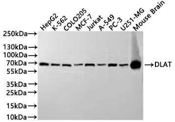
Western Blot
Positive WB detected in: HepG2 whole cell lysate(30µg), K562 whole cell lysate(30µg), COLO205 whole cell lysate(30µg), MCF7 whole cell lysate(30µg), Jurkat whole cell lysate(30µg), A549 whole cell lysate(30µg), PC-3 whole cell lysate(30µg), U251 whole cell lysate(30µg), Mouse brain tissue lysate(30µg)
All lanes:DLAT antibody at 1:1000
Secondary
Goat polyclonal to rabbit IgG at 1/40000 dilution
Predicted band size: 69 kDa
Observed band size: 69 kDa
Exposure time:2min
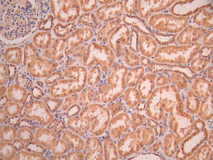
IHC image of CSB-RA283972A0HU diluted at 1:100 and staining in paraffin-embedded human kidney tissue performed on a Leica BondTM system. After dewaxing and hydration, antigen retrieval was mediated by high pressure in a citrate buffer (pH 6.0). Section was blocked with 10% normal goat serum 30min at RT. Then primary antibody (1% BSA) was incubated at 4°C overnight. The primary is detected by a Goat anti-rabbit polymer IgG labeled by HRP and visualized using 0.05% DAB.
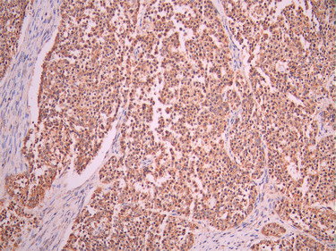
IHC image of CSB-RA283972A0HU diluted at 1:100 and staining in paraffin-embedded human liver cancer performed on a Leica BondTM system. After dewaxing and hydration, antigen retrieval was mediated by high pressure in a citrate buffer (pH 6.0). Section was blocked with 10% normal goat serum 30min at RT. Then primary antibody (1% BSA) was incubated at 4°C overnight. The primary is detected by a Goat anti-rabbit polymer IgG labeled by HRP and visualized using 0.05% DAB.
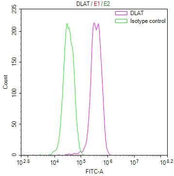
Overlay Peak curve showing MCF-7 cells stained with CSB-RA283972A0HU (red line) at 1:100. The cells were fixed in 4% formaldehyde and permeated by 0.2% TritonX-100 for 10min. Then 10% normal goat serum to block non-specific protein-protein interactions followed by the antibody (1ug/1*106cells) for 45min at 4℃. The secondary antibody used was FITC-conjugated goat anti-rabbit IgG (H+L) at 1/200 dilution for 35min at 4℃.Control antibody (green line) was Rabbit IgG (1ug/1*106cells) used under the same conditions. Acquisition of >10, 000 events was performed.
DLAT Antibody (CSB-PA006926LA01HU)
Validated Data
Western Blot
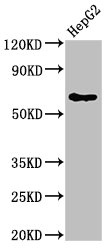
Positive WB detected in: HepG2 whole cell lysate
All lanes: DLAT antibody at 4μg/ml
Secondary
Goat polyclonal to rabbit IgG at 1/50000 dilution
Predicted band size: 69 kDa
Observed band size: 69 kDa
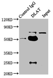
Immunoprecipitating DLAT in HepG2 whole cell lysate
Lane 1: Rabbit control IgG instead of CSB-PA006926LA01HU in HepG2 whole cell lysate.
For western blotting, a HRP-conjugated Protein G antibody was used as the secondary antibody (1/2000)
Lane 2: CSB-PA006926LA01HU (6μg) + HepG2 whole cell lysate (1mg)
Lane 3: HepG2 whole cell lysate (20μg)
The following DLAT reagents supplied by CUSABIO are manufactured under a strict quality control system. Multiple applications have been validated and solid technical support is offered.
DLAT Antibodies for Homo sapiens (Human)
| Code | Product Name | Species Reactivity | Application |
|---|---|---|---|
| CSB-PA006926GA01HU | DLAT Antibody | Human,Mouse,Rat | ELISA,WB |
| CSB-PA445587 | DLAT Antibody | Human,Mouse,Rat | ELISA,WB,IHC |
| CSB-PA084734 | DLAT Antibody | Human,Mouse,Rat | ELISA,WB,IHC |
| CSB-PA006926LA01HU | DLAT Antibody | Human | ELISA, WB, IP |
| CSB-RA283972A0HU | DLAT Recombinant Monoclonal Antibody | Human, Mouse | ELISA, WB, IHC, FC |
DLAT Antibodies for Mus musculus (Mouse)
| Code | Product Name | Species Reactivity | Application |
|---|---|---|---|
| CSB-PA804374ZA01MO | Dlat Antibody | Mus musculus | ELISA, WB (ensure identification of antigen) |
DLAT Proteins for Homo sapiens (Human)
| Code | Product Name | Source |
|---|---|---|
| CSB-YP006926HU CSB-BP006926HU CSB-MP006926HU CSB-EP006926HU-B |
Recombinant Human Dihydrolipoyllysine-residue acetyltransferase component of pyruvate dehydrogenase complex, mitochondrial (DLAT), partial | Yeast Baculovirus Mammalian cell In Vivo Biotinylation in E.coli |
| CSB-EP006926HU | Recombinant Human Dihydrolipoyllysine-residue acetyltransferase component of pyruvate dehydrogenase complex, mitochondrial (DLAT), partial | E.coli |
| CSB-EP006926HUc7 | Recombinant Human Dihydrolipoyllysine-residue acetyltransferase component of pyruvate dehydrogenase complex, mitochondrial (DLAT), partial | E.coli |
DLAT Proteins for Mus musculus (Mouse)
| Code | Product Name | Source |
|---|---|---|
| CSB-YP804374MO | Recombinant Mouse Dihydrolipoyllysine-residue acetyltransferase component of pyruvate dehydrogenase complex, mitochondrial (Dlat) | Yeast |
| CSB-EP804374MO CSB-BP804374MO CSB-EP804374MO-B |
Recombinant Mouse Dihydrolipoyllysine-residue acetyltransferase component of pyruvate dehydrogenase complex, mitochondrial (Dlat) | E.coli Baculovirus In Vivo Biotinylation in E.coli |
| CSB-EP804374MOa0 | Recombinant Mouse Dihydrolipoyllysine-residue acetyltransferase component of pyruvate dehydrogenase complex, mitochondrial (Dlat) | E.coli |
| CSB-MP804374MO | Recombinant Mouse Dihydrolipoyllysine-residue acetyltransferase component of pyruvate dehydrogenase complex, mitochondrial (Dlat) | Mammalian cell |
| CSB-MP804374MOe1 | Recombinant Mouse Dihydrolipoyllysine-residue acetyltransferase component of pyruvate dehydrogenase complex, mitochondrial (Dlat) | Mammalian cell |

