Call us
301-363-4651 (Available 9 a.m. to 5 p.m. CST from Monday to Friday)
The CBS gene encodes cystathionine β-synthase (CBS), a core enzyme in methionine’s transsulfuration pathway. Common aliases include CBS enzyme or cystathionine synthase. It catalyzes serine and homocysteine condensation into cystathionine, a key step in homocysteine catabolism and cysteine biosynthesis, maintaining homocysteine homeostasis.
CBS dysfunction causes hyperhomocysteinemia, associated with cardiovascular diseases (atherosclerosis, stroke) and neurological disorders (Alzheimer’s, epilepsy). Vitamin B6 supplementation (CBS relies on pyridoxal phosphate, a B6 derivative) is a standard treatment. Recent research focuses on small-molecule activators to boost CBS activity for managing homocysteine-related pathologies.
Recombinant Human Cystathionine beta-synthase (CBS), partial (CSB-EP004589HU)
Validated Data
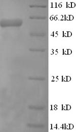
(Tris-Glycine gel) Discontinuous SDS-PAGE (reduced) with 5% enrichment gel and 15% separation gel.
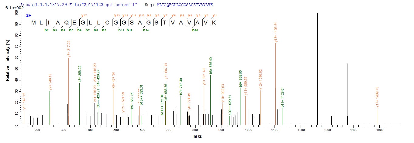
Based on the SEQUEST from database of E.coli host and target protein, the LC-MS/MS Analysis result of CSB-EP004589HU could indicate that this peptide derived from E.coli-expressed Homo sapiens (Human) CBS.
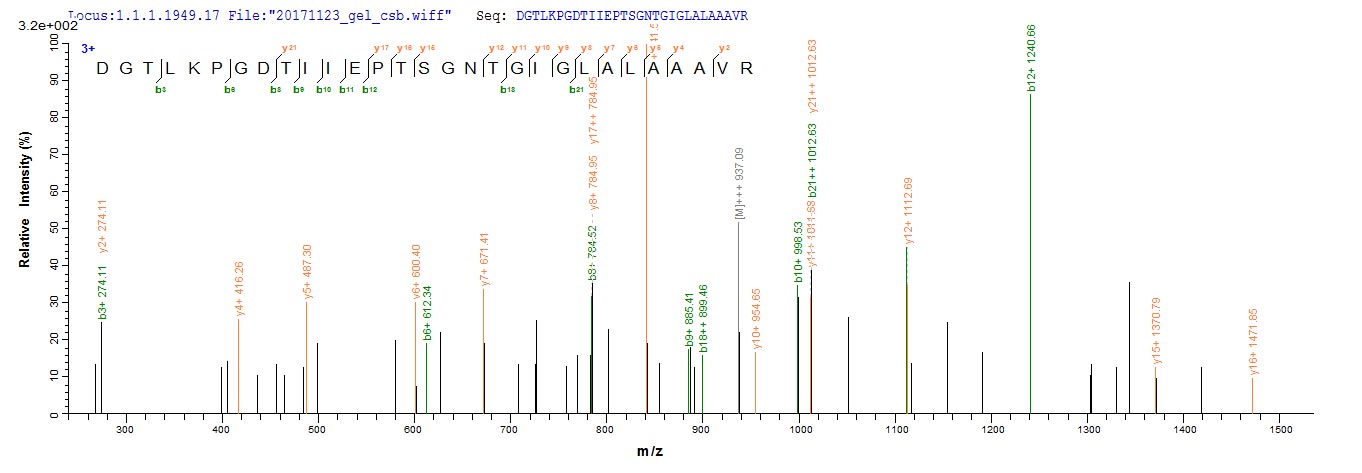
Based on the SEQUEST from database of E.coli host and target protein, the LC-MS/MS Analysis result of CSB-EP004589HU could indicate that this peptide derived from E.coli-expressed Homo sapiens (Human) CBS.
CBS Recombinant Monoclonal Antibody (CSB-RA242578A0HU)
Validated Data
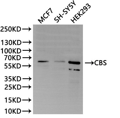
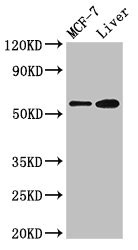
Western Blot
Positive WB detected in: MCF7 whole cell lysate(30µg), SH-SY5Y whole cell lysate(30µg), HEK293 whole cell lysate(30µg)
All lanes:CBS antibody at 1:1000
Secondary
Goat polyclonal to rabbit IgG at 1/40000 dilution
Predicted band size: 61 kDa
Observed band size: 61 kDa
Exposure time:2min
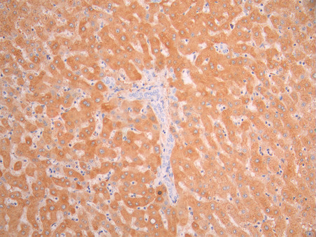
IHC image of CSB-RA242578A0HU diluted at 1:100 and staining in paraffin-embedded human liver tissue performed on a Leica BondTM system. After dewaxing and hydration, antigen retrieval was mediated by high pressure in a citrate buffer (pH 6.0). Section was blocked with 10% normal goat serum 30min at RT. Then primary antibody (1% BSA) was incubated at 4°C overnight. The primary is detected by a Goat anti-rabbit polymer IgG labeled by HRP and visualized using 0.05% DAB.
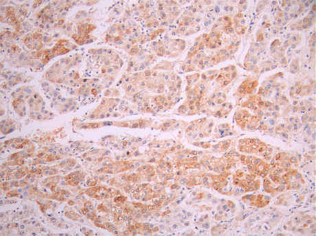
IHC image of CSB-RA242578A0HU diluted at 1:100 and staining in paraffin-embedded human liver cancer performed on a Leica BondTM system. After dewaxing and hydration, antigen retrieval was mediated by high pressure in a citrate buffer (pH 6.0). Section was blocked with 10% normal goat serum 30min at RT. Then primary antibody (1% BSA) was incubated at 4°C overnight. The primary is detected by a Goat anti-rabbit polymer IgG labeled by HRP and visualized using 0.05% DAB.
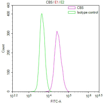
Overlay Peak curve showing SH-SY5Y cells stained with CSB-RA242578A0HU (red line) at 1:100. The cells were fixed in 4% formaldehyde and permeated by 0.2% TritonX-100 for 10min. Then 10% normal goat serum to block non-specific protein-protein interactions followed by the antibody (1ug/1*106cells) for 45min at 4℃. The secondary antibody used was FITC-conjugated goat anti-rabbit IgG (H+L) at 1/200 dilution for 35min at 4℃.Control antibody (green line) was Rabbit IgG (1ug/1*106cells) used under the same conditions. Acquisition of >10, 000 events was performed.
CBS Antibody (CSB-PA12719A0Rb)
Validated Data
Western Blot
Positive WB detected in: MCF-7 whole cell lysate, Mouse liver tissue
All lanes: CBS antibody at 2.7µg/ml
Secondary
Goat polyclonal to rabbit IgG at 1/50000 dilution
Predicted band size: 61, 62 kDa
Observed band size: 61 kDa
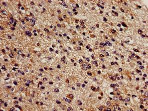
Immunohistochemistry analysis of human glioma using CSB-PA12719A0Rb at dilution of 1:100
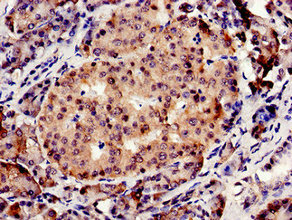
Immunocytochemistry analysis of human pancreatic tissue using CSB-PA12719A0Rb at dilution of 1:100
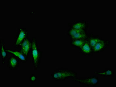
Immunofluorescent analysis of Hela cells using CSB-PA12719A0Rb at dilution of 1:100 and Alexa Fluor 488-congugated AffiniPure Goat Anti-Rabbit IgG(H+L)
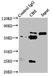
Immunoprecipitating CBS in Hela whole cell lysate
Lane 1: Rabbit control IgG instead of CSB-PA12719A0Rb in Hela whole cell lysate. For western blotting, a HRP-conjugated Protein G antibody was used as the secondary antibody (1/5000)
Lane 2: CSB-PA12719A0Rb (8µg) + Hela whole cell lysate (500µg)
Lane 3: Hela whole cell lysate (20µg)
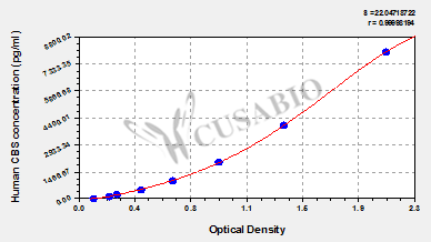
Human cystathionine β-synthase(CBS) ELISA Kit (CSB-E13314h)
Validated Data
Code: CSB-E13314h
Size: 96T,5×96T,10×96T
Sensitivity: 31.25 pg/mL
Detection Range: 125 pg/mL-8000 pg/mL
These standard curves are provided for demonstration only. A standard curve should be generated for each set of samples assayed.
The following CBS reagents supplied by CUSABIO are manufactured under a strict quality control system. Multiple applications have been validated and solid technical support is offered.
CBS Antibodies for Homo sapiens (Human)
| Code | Product Name | Species Reactivity | Application |
|---|---|---|---|
| CSB-PA004589GA01HU | CBS Antibody | Human,Mouse,Rat | ELISA,WB,IHC,IF |
| CSB-PA205503 | CBS Antibody | Human,Mouse,Rat | ELISA,WB,IHC |
| CSB-PA12719A0Rb | CBS Antibody | Human, Mouse | ELISA, WB, IHC, IF, IP |
| CSB-RA242578A0HU | CBS Recombinant Monoclonal Antibody | Human | ELISA, WB, IHC, FC |
CBS Proteins for Homo sapiens (Human)
| Code | Product Name | Source |
|---|---|---|
| CSB-YP004589HU | Recombinant Human Cystathionine beta-synthase (CBS), partial,Yeast | Yeast |
| CSB-EP004589HU | Recombinant Human Cystathionine beta-synthase (CBS), partial | E.coli |
| CSB-BP004589HU CSB-MP004589HU CSB-EP004589HU-B |
Recombinant Human Cystathionine beta-synthase (CBS), partial | Baculovirus Mammalian cell In Vivo Biotinylation in E.coli |
| CSB-BP004589HU1 CSB-MP004589HU1 CSB-EP004589HU1-B |
Recombinant Homo sapiens Cystathionine beta-synthasepartial | Baculovirus Mammalian cell In Vivo Biotinylation in E.coli |
| CSB-EP004589HU1 | Recombinant Human Cystathionine beta-synthase (CBS), partial | E.coli |
| CSB-YP004589HU1 | Recombinant Human Cystathionine beta-synthase (CBS), partial | Yeast |
CBS ELISA Kit for Homo sapiens (Human)
| Code | Product Name | Sample Types | Sensitivity |
|---|---|---|---|
| CSB-E13314h | Human cystathionine β-synthase(CBS) ELISA Kit | serum, plasma, tissue homogenates, cell lysates | 31.25 pg/mL |

