env Antibody

Rare Species
Code CSB-PA321953XA01GCA
Size US$299
Image
  • Western Blot
    Positive WB detected in: recombinant protein
    All lanes:env Antibody at 1:1000
    Secondary
    Goat polyclonal to rabbit IgG at 1/50000 dilution
    Predicted band size: 29 kDa
    Observed band size: 29 kDa
Promotion Free Antibody trial simple
Have Questions? Leave a Message or Start an on-line Chat

Product Details

Full Product Name
Rabbit anti-Gibbon ape leukemia virus (GALV) env Antibody Polyclonal antibody
Uniprot No.
Target Names
env Antibody
Alternative Names
Envelope glycoprotein (Env polyprotein) [Cleaved into: Surface protein (SU) (Glycoprotein 70) (gp70); Transmembrane protein (TM) (Envelope protein p15E); R-peptide (p2E)] env
Raised in
Rabbit
Species Reactivity
Gibbon ape leukemia virus (GALV)
Immunogen
Recombinant Gibbon ape leukemia virus (GALV) env protein (51-270)
Immunogen Species
Gibbon ape leukemia virus (GALV)
Conjugate
Non-conjugated
Clonality
Polyclonal
Isotype
IgG
Purification Method
>95%, Protein G purified
Concentration
It differs from different batches. Please contact us to confirm it.
Buffer
Preservative: 0.03% Proclin 300
Constituents: 50% Glycerol, 0.01M PBS, pH 7.4
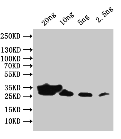
Form
Liquid
Tested Applications
ELISA, WB
Recommended Dilution
Application Recommended Dilution
WB 1:500-1:5000
Troubleshooting and FAQs
Storage
Upon receipt, store at -20°C or -80°C. Avoid repeated freeze.
Lead Time
Basically, we can dispatch the products out in 1-3 working days after receiving your orders. Delivery time maybe differs from different purchasing way or location, please kindly consult your local distributors for specific delivery time.
Usage
For Research Use Only. Not for use in diagnostic or therapeutic procedures.

Customer Reviews and Q&A

 Customer Reviews

There are currently no reviews for this product.

Submit a Review here

Target Background

Function
The surface protein (SU) attaches the virus to the host cell by binding to its receptor. This interaction triggers the refolding of the transmembrane protein (TM) and is thought to activate its fusogenic potential by unmasking its fusion peptide. Fusion occurs at the host cell plasma membrane.; The transmembrane protein (TM) acts as a class I viral fusion protein. Under the current model, the protein has at least 3 conformational states: pre-fusion native state, pre-hairpin intermediate state, and post-fusion hairpin state. During viral and target cell membrane fusion, the coiled coil regions (heptad repeats) assume a trimer-of-hairpins structure, positioning the fusion peptide in close proximity to the C-terminal region of the ectodomain. The formation of this structure appears to drive apposition and subsequent fusion of viral and target cell membranes. Membranes fusion leads to delivery of the nucleocapsid into the cytoplasm.
Subcellular Location
[Transmembrane protein]: Virion membrane; Single-pass type I membrane protein. Host cell membrane; Single-pass type I membrane protein.; [Surface protein]: Virion membrane; Peripheral membrane protein. Host cell membrane; Peripheral membrane protein.; [R-peptide]: Host cell membrane; Peripheral membrane protein.
Database Links

KEGG: vg:1491895

icon of phone
Call us
301-363-4651 (Available 9 a.m. to 5 p.m. CST from Monday to Friday)
icon of address
Address
7505 Fannin St., Ste 610, Room 7 (CUBIO Innovation Center), Houston, TX 77054, USA
icon of social media
Join us with

Subscribe newsletter

Leave a message

* To protect against spam, please pass the CAPTCHA test below.
CAPTCHA verification
© 2007-2025 CUSABIO TECHNOLOGY LLC All rights reserved. 鄂ICP备15011166号-1