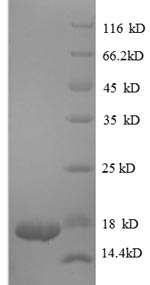
The creation of recombinant human TGFB1 protein begins by engineering an expression vector carrying the TGFB1 gene fragment (281-390aa) with the N-terminal 6xHis-tag gene, which is transfected into E.coli cells. Once the cells produce the protein, the purification process follows, often involving affinity chromatography to selectively bind and isolate the target protein. After purification, the protein’s purity is determined via SDS-PAGE, reaching above 90%.
The human TGFB1 protein is a multifunctional cytokine that plays critical roles in various biological processes, including cell growth, differentiation, immune regulation, and tissue repair [1]. TGFB1 is primarily involved in wound healing and tissue repair. It regulates the expression of extracellular matrix (ECM) proteins, angiogenic factors, and proteins that facilitate cell proliferation and migration, which are essential for the healing process [2]. Additionally, TGFB1 is a key player in fibrosis and scar formation, as it promotes the differentiation of fibroblasts into myofibroblasts, which are responsible for synthesizing ECM components such as collagen [3]. This fibrogenic activity is particularly relevant in conditions such as idiopathic pulmonary fibrosis, where TGFB1 signaling contributes to excessive ECM deposition and tissue remodeling [4].
Moreover, TGFB1 participates in immune system regulation. It modulates the proliferation and differentiation of various immune cells, including T cells and macrophages, and plays a role in maintaining immune tolerance [5]. The protein's immunosuppressive properties have made it a target for therapeutic strategies in cancer treatment, where it can promote tumor progression by creating an immunosuppressive microenvironment [6]. The signaling mechanisms of TGFB1 involve binding to specific receptors, which activate downstream signaling pathways, including the SMAD family of proteins that mediate its effects on gene expression [7].
References:
[1] F. Norozian, M. Kashyap, et al. Tgfβ1 induces mast cell apoptosis, Experimental Hematology, vol. 34, no. 5, p. 579-587, 2006. https://doi.org/10.1016/j.exphem.2006.02.003
[2] H. Madhyastha, S. Halder, et al. Surface refined auquercetinnanoconjugate stimulates dermal cell migration: possible implication in wound healing, RSC Advances, vol. 10, no. 62, p. 37683-37694, 2020. https://doi.org/10.1039/d0ra06690g
[3] P. Occhetta, G. Isu, et al. A three-dimensional in vitro dynamic micro-tissue model of cardiac scar formation, Integrative Biology, vol. 10, no. 3, p. 174-183, 2018. https://doi.org/10.1039/c7ib00199a
[4] A. Chen, Z. Sun, et al. Integrative bioinformatics and validation studies reveal kdm6b and its associated molecules as crucial modulators in idiopathic pulmonary fibrosis, Frontiers in Immunology, vol. 14, 2023. https://doi.org/10.3389/fimmu.2023.1183871
[5] I. Hayat, I. Lys, & R. Page. Novel network exploration through mrna changes in the skeletal muscle after resistance exercise, Pakistan Postgraduate Medical Journal, vol. 28, no. 1, p. 2-6, 2018. https://doi.org/10.51642/ppmj.v28i1.94
[6] N. Díaz-Valdés, M. Basagoiti, et al. Induction of monocyte chemoattractant protein-1 and interleukin-10 by tgfβ1 in melanoma enhances tumor infiltration and immunosuppression, Cancer Research, vol. 71, no. 3, p. 812-821, 2011. https://doi.org/10.1158/0008-5472.can-10-2698
[7] E. Lin, P. Kuo, Y. Liu, A. Yang, & S. Tsai. Transforming growth factor-β signaling pathway-associated genes smad2 and tgfbr2 are implicated in metabolic syndrome in a taiwanese population, Scientific Reports, vol. 7, no. 1, 2017. https://doi.org/10.1038/s41598-017-14025-4


