Call us
301-363-4651 (Available 9 a.m. to 5 p.m. CST from Monday to Friday)
| Code | CSB-RA569311A0HU |
| Size | US$210 |
| Order now | |
| Image |
|
| Have Questions? | Leave a Message or Start an on-line Chat |
| Application | Recommended Dilution |
|---|---|
| IF | 1:20-1:200 |
CXCR4, also known as CD184 or fusin, is a seven-transmembrane G protein-coupled receptor that serves as the primary receptor for stromal cell-derived factor 1 (SDF-1/CXCL12). This chemokine receptor plays a central role in leukocyte trafficking, hematopoietic stem cell homing, and embryonic development. Beyond its physiological functions, CXCR4 has garnered significant research attention for its involvement in cancer metastasis, where the CXCR4-SDF-1 axis guides tumor cell migration to secondary sites, and in HIV infection, where it functions as a co-receptor for viral entry.
This recombinant monoclonal antibody, clone 2F10, was generated against a synthetic peptide derived from human CXCR4 and produced in rabbit host. The recombinant format ensures consistent performance across experiments, eliminating the lot-to-lot variability that can complicate long-term studies or multi-site collaborations. With a sequence-defined binding region, you can confidently compare results over time and across different experimental setups.
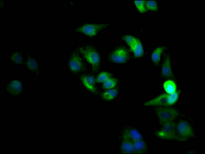
The antibody has been validated for immunofluorescence applications, where it successfully detected CXCR4 in HeLa cells at dilutions ranging from 1:20 to 1:200. In these experiments, cells were fixed with formaldehyde, permeabilized with Triton X-100, and visualized using FITC-conjugated secondary antibodies, demonstrating clear membrane and cytoplasmic staining patterns consistent with CXCR4 localization.
This antibody supports investigations across multiple disciplines, including cancer biology, immunology, neuroscience, stem cell research, and microbiology. Whether you are studying chemokine-mediated cell migration, characterizing tumor microenvironments, or investigating stem cell trafficking mechanisms, this recombinant antibody provides the reliability needed for reproducible immunofluorescence-based detection of human CXCR4.
Applications : Immunohistochemistry (IHC)
Sample type: cell
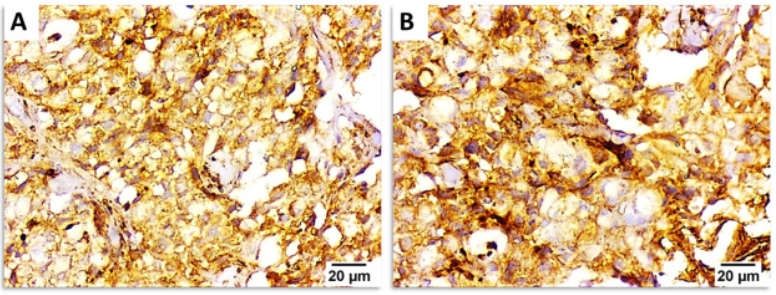
Review: Representative photomicrographs for immunohistochemical staining of CXCR4. Invasive ductal carcinoma showing moderate [A] and strong [B] cytoplasmic and membranous staining of CXCR4. Brown color indicates positive staining. Negative, weak, moderate and strong staining = 0, 1, 2 and 3, respectively. Magnification: ×400, scale bar: 20 μm.
By Anonymous

