MAPK3 Antibody

Code CSB-PA013456LA01HU
Size US$166
Order now
Image
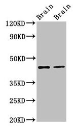
  • Western Blot
    Positive WB detected in: Rat brain tissue, Mouse brain tissue
    All lanes: MAPK3 antibody at 3µg/ml
    Secondary
    Goat polyclonal to rabbit IgG at 1/50000 dilution
    Predicted band size: 44, 39, 41 kDa
    Observed band size: 44 kDa

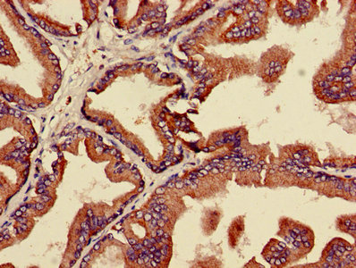
  • IHC image of CSB-PA013456LA01HU diluted at 1:600 and staining in paraffin-embedded human prostate cancer performed on a Leica BondTM system. After dewaxing and hydration, antigen retrieval was mediated by high pressure in a citrate buffer (pH 6.0). Section was blocked with 10% normal goat serum 30min at RT. Then primary antibody (1% BSA) was incubated at 4°C overnight. The primary is detected by a biotinylated secondary antibody and visualized using an HRP conjugated SP system.

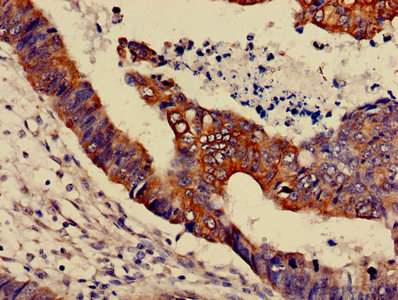
  • IHC image of CSB-PA013456LA01HU diluted at 1:600 and staining in paraffin-embedded human colon cancer performed on a Leica BondTM system. After dewaxing and hydration, antigen retrieval was mediated by high pressure in a citrate buffer (pH 6.0). Section was blocked with 10% normal goat serum 30min at RT. Then primary antibody (1% BSA) was incubated at 4°C overnight. The primary is detected by a biotinylated secondary antibody and visualized using an HRP conjugated SP system.

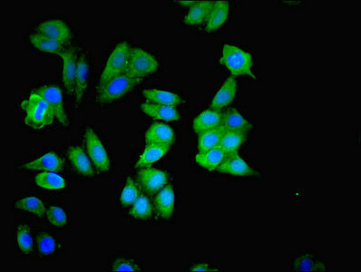
  • Immunofluorescence staining of HepG2 cells with CSB-PA013456LA01HU at 1:200, counter-stained with DAPI. The cells were fixed in 4% formaldehyde, permeabilized using 0.2% Triton X-100 and blocked in 10% normal Goat Serum. The cells were then incubated with the antibody overnight at 4°C. The secondary antibody was Alexa Fluor 488-congugated AffiniPure Goat Anti-Rabbit IgG(H+L).

The Latest Promotion Free Antibody trial simple
Have Questions? Leave a Message or Start an on-line Chat

Product Details

Full Product Name
Rabbit anti-Homo sapiens (Human) MAPK3 Polyclonal antibody
Uniprot No.
Target Names
MAPK3
Alternative Names
ERK 1 antibody; ERK-1 antibody; ERK1 antibody; ERT 2 antibody; ERT2 antibody; Extracellular Signal Regulated Kinase 1 antibody; Extracellular signal related kinase 1 antibody; Extracellular signal-regulated kinase 1 antibody; HGNC6877 antibody; HS44KDAP antibody; HUMKER1A antibody; Insulin Stimulated MAP2 Kinase antibody; Insulin-stimulated MAP2 kinase antibody; MAP kinase 1 antibody; MAP kinase 3 antibody; MAP Kinase antibody; MAP kinase isoform p44 antibody; MAPK 1 antibody; MAPK 3 antibody; MAPK antibody; MAPK1 antibody; Mapk3 antibody; MGC20180 antibody; Microtubule Associated Protein 2 Kinase antibody; Microtubule-associated protein 2 kinase antibody; Mitogen Activated Protein Kinase 3 antibody; Mitogen-activated protein kinase 1 antibody; Mitogen-activated protein kinase 3 antibody; MK03_HUMAN antibody; OTTHUMP00000174538 antibody; OTTHUMP00000174541 antibody; p44 ERK1 antibody; p44 MAPK antibody; p44-ERK1 antibody; p44-MAPK antibody; P44ERK1 antibody; P44MAPK antibody; PRKM 3 antibody; PRKM3 antibody; Protein Kinase Mitogen Activated 3 antibody
Raised in
Rabbit
Species Reactivity
Human, Mouse, Rat
Immunogen
Recombinant Human Mitogen-activated protein kinase 3 protein (11-379AA)
Immunogen Species
Homo sapiens (Human)
Conjugate
Non-conjugated

The MAPK3 Antibody (Product code: CSB-PA013456LA01HU) is Non-conjugated. For MAPK3 Antibody with conjugates, please check the following table.

Available Conjugates
Conjugate Product Code Product Name Application
HRP CSB-PA013456LB01HU MAPK3 Antibody, HRP conjugated ELISA
FITC CSB-PA013456LC01HU MAPK3 Antibody, FITC conjugated
Biotin CSB-PA013456LD01HU MAPK3 Antibody, Biotin conjugated ELISA
Clonality
Polyclonal
Isotype
IgG
Purification Method
>95%, Protein G purified
Concentration
It differs from different batches. Please contact us to confirm it.
Buffer
Preservative: 0.03% Proclin 300
Constituents: 50% Glycerol, 0.01M PBS, PH 7.4
Form
Liquid
Tested Applications
ELISA, WB, IHC, IF
Recommended Dilution
Application Recommended Dilution
WB 1:500-1:5000
IHC 1:500-1:1000
IF 1:200-1:500
Troubleshooting and FAQs
Storage
Upon receipt, store at -20°C or -80°C. Avoid repeated freeze.
Lead Time
Basically, we can dispatch the products out in 1-3 working days after receiving your orders. Delivery time maybe differs from different purchasing way or location, please kindly consult your local distributors for specific delivery time.

Customer Reviews and Q&A

 Customer Reviews
Average Rating:
5.0 - 1 reviews

Submit a Review here

Applications : WB

Sample dilution: 1: 1000

Review: Western blot analysis of total ERK (t-ERK 1/2), p-ERK 1/2 and GAPDH in DRG of mice injected with/without CaP.

By Anonymous

Target Background

Function
Serine/threonine kinase which acts as an essential component of the MAP kinase signal transduction pathway. MAPK1/ERK2 and MAPK3/ERK1 are the 2 MAPKs which play an important role in the MAPK/ERK cascade. They participate also in a signaling cascade initiated by activated KIT and KITLG/SCF. Depending on the cellular context, the MAPK/ERK cascade mediates diverse biological functions such as cell growth, adhesion, survival and differentiation through the regulation of transcription, translation, cytoskeletal rearrangements. The MAPK/ERK cascade plays also a role in initiation and regulation of meiosis, mitosis, and postmitotic functions in differentiated cells by phosphorylating a number of transcription factors. About 160 substrates have already been discovered for ERKs. Many of these substrates are localized in the nucleus, and seem to participate in the regulation of transcription upon stimulation. However, other substrates are found in the cytosol as well as in other cellular organelles, and those are responsible for processes such as translation, mitosis and apoptosis. Moreover, the MAPK/ERK cascade is also involved in the regulation of the endosomal dynamics, including lysosome processing and endosome cycling through the perinuclear recycling compartment (PNRC); as well as in the fragmentation of the Golgi apparatus during mitosis. The substrates include transcription factors (such as ATF2, BCL6, ELK1, ERF, FOS, HSF4 or SPZ1), cytoskeletal elements (such as CANX, CTTN, GJA1, MAP2, MAPT, PXN, SORBS3 or STMN1), regulators of apoptosis (such as BAD, BTG2, CASP9, DAPK1, IER3, MCL1 or PPARG), regulators of translation (such as EIF4EBP1) and a variety of other signaling-related molecules (like ARHGEF2, FRS2 or GRB10). Protein kinases (such as RAF1, RPS6KA1/RSK1, RPS6KA3/RSK2, RPS6KA2/RSK3, RPS6KA6/RSK4, SYK, MKNK1/MNK1, MKNK2/MNK2, RPS6KA5/MSK1, RPS6KA4/MSK2, MAPKAPK3 or MAPKAPK5) and phosphatases (such as DUSP1, DUSP4, DUSP6 or DUSP16) are other substrates which enable the propagation the MAPK/ERK signal to additional cytosolic and nuclear targets, thereby extending the specificity of the cascade.
Gene References into Functions
  1. Data suggest that Thr264 in TRPV3 is key ERK1 phosphorylation site mediating EGFR-induced sensitization of TRPV3 to stimulate signaling pathways involved in regulating skin homeostasis. (TRPV3 = transient receptor potential cation channel subfamily V member-3; ERK1 = extracellular signal-regulated kinase-1; EGFR = epidermal growth factor receptor) PMID: 29084846
  2. RASSF7 promotes cell proliferation through activating MEK1/MEK2-ERK1/ERK2 signaling pathway in hepatocellular carcinoma. PMID: 29729697
  3. ERK1 Directly Interacts With JNK1 Leading to Regulation of JNK1/c-Jun Activity and Cell Transformation. PMID: 28106280
  4. the D domain of LRRC4 anchors ERK1/2 in the cytoplasm and competitively inhibits MEK/ERK activation in glioma cells. PMID: 27884160
  5. High ERK1 expression is associated with castration-resistant prostate cancer. PMID: 28844715
  6. The antitumor activity of scopoletin may be due to its strong anti-angiogenic effect, which may be mediated by its effective inhibition of ERK1, VEGF-A, and FGF-2. PMID: 27133199
  7. High ERK1 expression is associated with melanoma. PMID: 28193911
  8. findings uncover a role of ERK1 in the regulation of furin activity by supporting a self-sustaining loop for high TGF-beta activity in glioma-initiating cells. PMID: 28484053
  9. ERK1 phosphorylation is mediated by Src and Csk. PMID: 26234813
  10. Integrin beta1 appears to serve as a partner of Stathmin induction of ERK and Akt signaling by inhibiting apoptosis in the cholangiocarcinoma cell. PMID: 28178656
  11. High ERK1 expression is associated with gastric cancer. PMID: 27601158
  12. Data indicate three biomarkers mitogen-activated protein kinase 3 (MAPK3), BCL2 apoptosis regulator (BCL2) and proto-oncogene c-Akt (AKT1) as potential predictors of neurological outcome following cardiac arrest (CA). PMID: 28147324
  13. Inhibiting miR-21 attenuates hepatic fibrosis by suppressing both the ERK1 in hepatic stellate cells and epithelial-mesenchymal transition of hepatocyte. PMID: 27226339
  14. High ERK1 expression is associated with paclitaxel resistance in cervical cancer. PMID: 26810068
  15. High ERK1 expression is Associated with Nucleus Pulposus Cells' Degeneration. PMID: 27635110
  16. Low expression of ERK is associated with resistance to sorafenib in liver cancer. PMID: 26711788
  17. Increased ERK1 expression is associated with drug resistance in neoplasms. PMID: 26715278
  18. The activation of ERK1 by MEK1, subsequent slower phosphorylation of the flanking sites results in inhibition of the kinase. Because the T207 and Y210 phosphosites of ERK1 are highly conserved within the eukaryotic protein kinase family, hyperphosphorylation within the kinase activation T-loop may serve as a general mechanism for protein kinase down-regulation after initial activation by their upstream kinases. PMID: 26823016
  19. Over-expressed TWIST associates with markers of epithelial mesenchymal transition and predicts poor prognosis in breast cancers via ERK and AKT activation. PMID: 26295469
  20. p44/42, a known apoptosis-promoting regulator and caspase 3 activator, was increased in brain tumor cells treated with violacein PMID: 25816226
  21. miR-155 plays an important role in regulating the pathological network involving EMT process and ERK1 pathway during hepatic stellate cell activation. PMID: 25142507
  22. This study identified and confirmed MAPK3 protein changes within the postsynaptic density in schizophrenia PMID: 25048004
  23. Sphingosine-1-phosphate promotes extravillous trophoblast cell invasion by activating MEK/ERK/MMP-2 signaling pathways via S1P/S1PR1 axis activation. PMID: 25188412
  24. Results show that miR-483-5p expression level is up-regulated in polycystic ovary syndrome patients and inversely correlated with notch3 and MAPk3 levels and that Notch3 and MAPK3 are the direct targets of MIR483. PMID: 25622783
  25. SKLB-M8 inhibited HUVEC proliferation, migration, invasion, and tube formation in vitro with the inhibition of phosphorylated ERK1/2. PMID: 25341684
  26. Therefore, the positivity of p-ERK1/2 expression may serve as a vital biomarker in the development of non-small cell lung cancer PMID: 25596700
  27. Low ERK1 expression is associated with hormone resistance in breast cancer. PMID: 25085753
  28. Therefore, our study demonstrates that MAPK members (ERK1/2 and JNK) play a key role in CCR7 regulating SCCHN metastasis PMID: 25270024
  29. High phosphorylated ERK1 is associated with low response to chemotherapy in nonsmall-cell lung carcinoma. PMID: 25449334
  30. These results therefore indicate that p53-mediated up-regulation of MKP-3 contributes to the establishment of the senescent cellular phenotype through dephosphorylating ERK1/2 PMID: 25414256
  31. ERK1/2 signal induced MNK catalytic activity enabled enterovirus type 1 internal ribosomal entry site-mediated translation/host cell cytotoxicity through negative regulation of the Ser/Arg (SR)-rich protein kinase (SRPK). PMID: 25187541
  32. ERK, AKT, and GSK-3beta have roles in boldine-induced cell cycle arrest and apoptosis in T24 human bladder cancer cell line PMID: 24239461
  33. Insulin-induced apoptotic commitment depended on the down-regulation of Erk-1, insulin growth factor-1 receptor (IGF-1R), and fibroblast growth factor receptor-1 (FGFR-1)-mediated signaling. PMID: 24818995
  34. Enhanced t-ERK1 expression in infiltrating lymphoid cells was significantly associated with female gender, absence of vascular and perineural invasion, lymph node metastases and early depth of invasion as well as with longer disease-free survival times PMID: 24682903
  35. Expression of TMPRSS4 in gastric cancer is significantly associated with lymph node and distant metastasis, high Erk1 expression, and poor prognosis. PMID: 23922976
  36. ERK1 phosphorylates KIBRA at Ser(548) for cell proliferation and migratory activity PMID: 24269383
  37. We conclude that pERK1/2 is a sensitive marker of early colon cancer, which disappears at later stages of cancer development. PMID: 23357054
  38. In activated eosinophils ligation of Siglec-8 leads to ROS-dependent enhancement of IL-5-induced ERK phosphorylation, which results in a novel mode of biochemically regulated eosinophil cell death. PMID: 23684072
  39. ERK1 localized to the cytosol and translocated to the nucleus upon cell activation and kinase phosphorylation. PMID: 23651922
  40. Data indicate that tocilizumab enhanced the interferon-induced phosphorylation of STAT1 and inhibited SOCS3 expression and the phosphorylation of both STAT3 and ERK. PMID: 23274199
  41. Extracellular signal-regulated kinase and glycogen synthase kinase 3beta regulate gephyrin postsynaptic aggregation and GABAergic synaptic function in a calpain-dependent mechanism PMID: 23408424
  42. ERK1/2 signaling plays an important role in topoisomerase II poison-induced G2/M checkpoint activation. PMID: 23166842
  43. MRK is a novel RhoC effector that controls LPA-stimulated cell invasion at least in part by regulating myosin dynamics, ERK and p38 PMID: 23319595
  44. osteosarcoma patients whose tumors expressed pERK1 had a poorer clinical outcome than those whose tumors did not. PMID: 22935974
  45. Results showed significantly higher levels of ERK1 protein in smokers vs. non-smokers. Analysis revealed a significant relation among the number of cigarettes smoked daily, the Fagerstrom Test for Nicotine Dependence score and the mRNA expression of ERK1. PMID: 21070506
  46. constitutive activation of the ERK1 pathway in HER2/ERBB2-transformed cells prevents EGF deprivation-induced FLIPL upregulation and TRAIL resistance. PMID: 22722337
  47. Lead specifically induces dysregulation of iron response element (IRP)1 protein by activating the ERK1/2 signaling pathway, indicating a novel role for IRP1 and the ERK/MAPK pathway in vascular endothelial functions. PMID: 22502979
  48. CXCL12/CXCR4 protein signaling axis induces sonic hedgehog expression in pancreatic cancer cells via extracellular regulated kinase- and Akt kinase-mediated activation of nuclear factor kappaB PMID: 22995914
  49. Aortic endothelial cells stimulated with HLA class I antibodies did not promote any detectable change in intracellular Ca(2+) concentration but instead induced MLC phosphorylation and stress fiber assembly. PMID: 22914643
  50. The inhibition of Id-1 expression by MK615 is mediated via ERK1/2 activation. PMID: 22076920

Show More

Hide All

Subcellular Location
Cytoplasm. Nucleus. Membrane, caveola. Cell junction, focal adhesion.
Protein Families
Protein kinase superfamily, CMGC Ser/Thr protein kinase family, MAP kinase subfamily
Database Links

HGNC: 6877

OMIM: 601795

KEGG: hsa:5595

STRING: 9606.ENSP00000263025

UniGene: Hs.861

CUSABIO guaranteed quality
icon of phone
Call us
301-363-4651 (Available 9 a.m. to 5 p.m. CST from Monday to Friday)
icon of address
Address
7505 Fannin St., Ste 610, Room 7 (CUBIO Innovation Center), Houston, TX 77054, USA
icon of social media
Join us with

Subscribe newsletter

Leave a message

* To protect against spam, please pass the CAPTCHA test below.
CAPTCHA verification
© 2007-2024 CUSABIO TECHNOLOGY LLC All rights reserved. 鄂ICP备15011166号-1
webinars: DT3C facilitates antibody internalization X
Place an order now

I. Product details

*
*
*
*

II. Contact details

*
*

III. Ship To

*
*
*
*
*
*
*

IV. Bill To

*
*
*
*
*
*
*
*