Call us
301-363-4651 (Available 9 a.m. to 5 p.m. CST from Monday to Friday)
What is autophagy? Autophagy is the natural, conserved degradation of the cell that plays a housekeeping role in removing misfolded or aggregated proteins, clearing damaged organelles, as well as eliminating intracellular pathogens through a lysosome-dependent regulated mechanism. It is a self-degradative process that is important for balancing sources of energy at critical times in development and in response to nutrient stress [1].
Generally speaking, the process of autophagy is divided into four stages:
Stage I: After the autophagy-inducing signal is received by the cells, a small membrane structure is formed in the cytoplasm. This structure is a bowl-like structure with a non-spherical, flat bilayer membrane and continues to expand. It is called an phagophore. This structure observed under electron microscopy is one of the gold standards for indicating the occurrence of autophagy.
Stage II: the continuously extending phagophore encapsulates several components (including organelles) in the cytoplasm together to form a closed spherical autophagosome. Autophagosome is characterized by double-layered membrane and containing cytoplasmic components (fragments of the endoplasmic reticulum and mitochondria, etc.).
Stage III: After autophagosome is formed, it may fuse with phagocyticvacuole, pinosome and endosome (this stage is a non-essential step).
Stage IV: autophagosome fuses with lysosome to form autolysosome. In this stage, lysosomal enzyme degrades the inner membrane of the autophagosome, so that the contents of the two are combined. Then, the contents in the autophagosome are degraded, and the products such as amino acids and fatty acids are transported to the cytoplasm and reused for supply, and the residue is excreted out of the cell or retained in the cytoplasm [2].
Based on different ways of intracellular substrates transported to the lysosome lumen, autophagy in mammalian is divided into three types, including macroautophagy, microautophagy and chaperone-mediated autophagy. Macroautophagy is the main form of autophagy. In macroautophagy, some membrane structures in the cytoplasm form autophagosomes by expanding to surround the degraded material, and then fuse with lysosomes and degrade their contents (as mentioned on the last paragraph). In microautophagy, the invagination of the membrane of the lysosome directly wraps the cell contents and degrades it in the lysosome. Microautophagy doesn’t have the stage of forming autophagosome. In chaperone-mediated autophagy (CMA), intracytoplasmic proteins bound to molecular chaperones are transported into the lysosomal lumen and then digested by lysosomal enzymes. Comparing with other two forms of autophagy, CMA is selective in scavenging proteins [3].
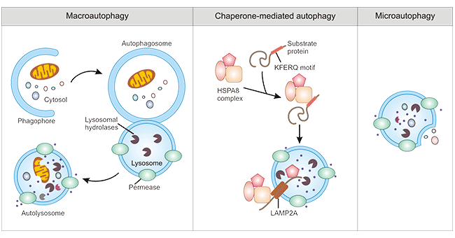
Figure 1. Three types of autophagy in mammalian
Additionally, according to the selectivity of autophagy to degrade substrates, autophagy can be further divided into mitophagy, reticulophagy, ribophagy, pexophagy, etc. Among of them, mitophagy is the most popular with researchers.
Autophagy is a cellular catabolic pathway involving in protein degradation, organelle turnover, and non-selective breakdown of cytoplasmic components. Autophagy initiation starts with the activation of the ULK1 complexes (including ULK1, ULK2, Atg13, RB1CC1, and Atg101), which activates different class III PI3K complexes composed of the core components (Beclin-1, VPS34/PIK3C3) and other recruited variable components (such as Atg14L, UVRAG, AMBRA1, etc.). These class III PI3K complexes play different functions [4] [5]. The Atg5-Atg12 complex bound to Atg16 mediates autophagosome membrane expansion, and LC3 and GABARAP family members bind to the lipid phosphatidylethanolamine (PE) and are recruited to the membrane. After Atg4 binds to Atg7, LC3-I and PE are coupled to form the lipid-bound form LC3-II, which is often used as a marker for autophagosomes [6]. more information of macroautophagy>>.
CUSABIO lists partial popular targets related autophagy, click to see all the related molecules/targets and research reagents of them.
References
[1] Danielle Glick, Sandra Barth, and Kay F. Macleod. Autophagy: cellular and molecular mechanisms [J]. J Pathol. 2010 May; 221(1): 3-12.
[2] Mizushima N. Autophagy: process and function [J]. Genes Dev. 2007, 21 (22): 2861-73.
[3] Parzych KR, Klionsky DJ. An overview ofautophagy: morphology, mechanism, and regulation [J]. Antioxid Redox Signal. 2014Jan 20;20(3):460-73.
[4] Zhiping Xie, Daniel J Klionsky. Autophagosome formation: core machinery and adaptations [J]. Nature Cell Biology. 2007, 9: 1102-1109.
[5] Jean M Mulcahy Levy, Christina G Towers, Andrew Thorburn. Targeting autophagy in cancer [J]. Nat Rev Cancer. 2017, 17(9): 528-542.
[6] Elena Shvets & Zvulun Elazar. Autophagy-independent incorporation of GFPLC3 into protein aggregates is dependent on its interaction with p62/SQSTM1 [J]. Autophagy. 2007, 3(4):323-8.
Popular Topics
Related Articles