Call us
301-363-4651 (Available 9 a.m. to 5 p.m. CST from Monday to Friday)
Before introducing ferroptosis, we learn something about iron in brief. Iron is an important trace element that maintains the life of organisms, and plays an important role in human oxidative metabolism, cell growth and proliferation, and oxygen transport and storage. About half of the enzymes and co-enzymes participating in the tricarboxylic acid cycle contain iron or require iron assistance. So what is Ferroptosis? Ferroptosis is a new type of cell death that is discovered by Stockwell in 2012. It is usually accompanied by a large amount of iron accumulation and lipid peroxidation during the cell death process. As the item implies, the occurrence of ferroptosis is iron-dependent. Ferroptosis has played important roles in multiple system diseases, including nervous system diseases, heart diseases, liver diseases, cancers, and so on [1].
Comparing with other types of cell death (such as apoptosis), ferroptosis is a novel type of cell death with distinct properties as follows:
Morphologically, it is characterized by the presence of smaller than normal mitochondria with condensed mitochondrial membrane densities, reduction or vanishing of mitochondria crista, and outer mitochondrial membrane rupture. Moreover, the cell membrane is ruptured and blebbed, and the nucleus is normal in size but lacks chromatin condensation.
Biologically, it is characterized by accumulation of intracellular iron and ROS, activation of mitogen-activated protein kinase (MAPK) signaling system, glutathione (GSH) consumption reduction, oxidation of nicotinamide adenine dinucleotide phosphate (NADPH) increase.
Ferroptosis is mainly caused by the imbalance between the production and degradation of intracellular lipid ROS. When the antioxidant capacity of cells is reduced, the accumulation of lipid reactive oxygen species can cause oxidative cell death, that is, ferroptosis.
The initiation and execution of ferroptosis are linked to the regulation of amino acid, lipid and iron metabolism and are regulated by multiple molecular mechanisms. These mechanisms can be roughly divided into three categories (As the figure 1 shows).
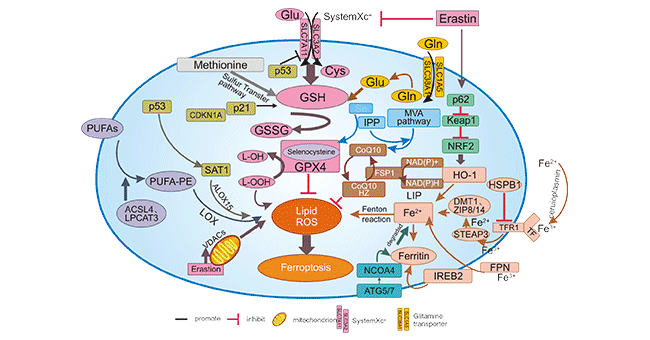
Figure 1. Regulatory pathways of ferroptosis
The first one, a major type, is regulated by GSH/GPX4 pathway, such as inhibition of system Xc-, inhibition of GPX4, sulfur transfer pathway, mevalonate (MVA) pathway, glutamine pathway, and p53 regulatory axis. System Xc-, a heterodimer composed of two subunits, SLC7A11 and SLC3A2, is an amino acid antitransporter widely distributed in phospholipid bilayers. SLC7A11 is the major functional subunit that transports cystine into the cell for GSH synthesis. Inhibition of SLCTA11 expression induces ferroptosis. As a tumor suppressor gene, p53 inhibits the uptake of cystine by cells through down-regulating the expression of SLC7A11 (a component of systemXC), resulting in decreased glutathione peroxidase activity, reduced cellular antioxidant capacity, and enhanced cellular response to ferroptosis. sensitivity.
The second one is the regulation mechanism of iron metabolism, including the regulation of ATG5-ATG7-NCOA4 pathway and IREB2 related to ferritin metabolism, and the regulatory pathways of p62-Keap1-NRF2 and HSPB1 [2] [3]. The last one is related pathways around lipid metabolism, such as P53-SAT1-ALOX15, ACSL4, LPCAT3, etc., which have effects on lipid regulation and ferroptosis. In addition, Erastin acts on mitochondria to induce ferroptosis [4] [5].
CUSABIO lists partial popular targets related ferroptosis, click to see all the related molecules/targets and research reagents of them.
References
[1] Li, J., Cao, F., Yin, Hl. et al. Ferroptosis: past, present and future [J]. Cell Death Dis. 2020, 11, 88.
[2] Sun, X. et al. HSPB1 as a novel regulator of ferroptotic cancer cell death [J]. Oncogene. 2015, 34, 5617–25.
[3] Gammella, E., Recalcati, S., Rybinska, I. et al. Iron-induced damage in cardiomyopathy: oxidative-dependent and independent mechanisms [J]. Oxid. Med Cell Longev. 2015, 230182.
[4] Yang, W. S. & Stockwell, B. R. Ferroptosis: death by lipid peroxidation [J]. Trends Cell Biol. 2016, 26, 165–176.
[5] Kagan, V. E. et al. Oxidized arachidonic and adrenic PEs navigate cells to ferroptosis [J]. Nat. Chem. Biol. 2017, 13, 81–90.
Popular Topics
Related Articles