Call us
301-363-4651 (Available 9 a.m. to 5 p.m. CST from Monday to Friday)
Anti-tag antibodies and epitope tags can form a stringent antigen-antibody-specific binding pair. Among these, epitope tags are short known peptides expressed through genetic engineering as fusions with target proteins. Anti-tag antibodies, serving as highly specific molecular probes, can recognize these short peptide sequences, thereby enabling qualitative, quantitative, localization, and affinity purification analyses of the target fusion proteins. In the field of bioengineering, the two together establish a standardized research platform. As a universal "identifier" fused to target proteins, epitope tags, in synergy with highly specific anti-tag antibodies, enable high-throughput detection and purification of various recombinant proteins. This system is widely applied in biopharmaceutical process monitoring, quality control, and protein interaction analysis in functional genomics, significantly enhancing research efficiency and standardization.
Epitope Tag, also known as tag peptides is a technical tool widely used in molecular biology, biochemistry, and cell biology research. It refers to the process of linking a known, short exogenous peptide sequence (typically 8-15 amino acids) to a target protein. N-terminal or C-terminalMan-madeFusion expression, thereby enabling the detection, purification, or localization of the target protein using specific antibodies against the tag. Epitope tags and fusion proteins.Significantly distinct:Fusion protein is a broad concept that refers to a protein expressed by linking two different genes together through DNA recombination technology (e. g., GFP fluorescent protein + target protein). In contrast, an epitope tag merely provides a "sequence recognized by an antibody" and does not possess biological activities such as catalysis or luminescence. Its primary function is to serve as a handle for detection and purification.
| Tag Type | Sequence and Size | Advantage | Limitations | Typical Application Scenarios |
|---|---|---|---|---|
| FLAG tag | Sequence: DYKDDDDK |
1. High Specificity: Short sequence design reduces non-specific binding, suitable for experiments such as immunoprecipitation (IP) and Western Blot. 2. AntibodyRich: For example, the M2 anti-FLAG® antibody (Sigma-Aldrich) has been extensively validated and supports a variety of detection methods. 3. Cleavability: Contains an enterokinase cleavage site, facilitating tag removal to obtain the native protein. |
1. Small molecular weight: may affect the native conformation or function of certain small proteins. 2. Negatively charged: May affect protein migration rate in electrophoresis, requiring optimization of experimental conditions. 3. Higher cost:AntibodyThe price is higher than some other tag antibodies. |
1. Protein-Protein Interaction Studies: Co-immunoprecipitation (IP) is used to capture target proteins and their interacting partners. 2. Drug Screening: Combining flow cytometry (FCM) to analyze changes in cell surface receptor expression. 3. Gene Therapy: Validation of the Purity and Activity of Viral Vector Packaging Proteins. |
| Size: 8 amino acids (approximately 1 kDa) | ||||
| HA tag | Sequence: YPYDVPDYA |
1. Natural Source: Derived from the hemagglutinin protein of the influenza virus, with low immunogenicity, suitable for live-cell imaging. 2. Multi-application scenarios: Supports Western Blot, IP, flow cytometry (FCM), and immunofluorescence (IF). 3. High-affinity antibodies: For example, the 12CA5 antibody (Roche) has high sensitivity and can detect low-abundance proteins. |
1. Longer sequence: May increase steric hindrance after protein fusion, affecting function. 2. Cross-reactivity Risk: Weak binding with certain proteins in host cells necessitates the inclusion of negative controls. 3. Low cleavage efficiency: The lack of high-efficiency restriction sites makes tag removal more difficult. |
1. Live Cell Dynamic Tracking: Immunofluorescence (IF) for observing protein subcellular localization (e. g., nucleocytoplasmic transport). 2. Vaccine Development: Detection of surface antigen expression on virus-like particles (VLPs). 3. Cell Signaling Pathways: Analysis of Endocytosis and Degradation of Receptor Tyrosine Kinases (RTKs). |
| Size: 9 amino acids (approximately 1 kDa) | ||||
| Myc tag | Sequence: EQKLISEEDL |
1. Classic Tag: Widely used for protein expression validation, interaction studies, and subcellular localization analysis. 2. Multi-Technology Compatibility: Supports Western Blot, IP, IF, FCM, and Chromatin Immunoprecipitation (ChIP). 3. High-sensitivity antibodies: For example, the 9E10 antibody (Santa Cruz) can detect nanogram-level proteins. |
1. Larger molecular weight: may affect the folding or activity of small proteins. 2. Charged Sequence: May interfere with the surface charge distribution of the protein, requiring evaluation of its impact on function. 3. Batch-to-Batch Variation: Some antibodies exhibit inter-batch differences.DifferenceExperimental conditions need to be optimized. |
1. Transcription Factor Studies: Chromatin Immunoprecipitation (ChIP) analysis of DNA binding activity. 2. Protein Stability: Monitor protein degradation rates through pulse-chase experiments. 3. Preclinical testing: ELISA quantitative detection of recombinant protein drug concentration in serum. |
| Size: 10 amino acids (approximately 1.2 kDa) | ||||
| V5 tag | Sequence: GKPIPNPLLGLDST |
1. Long Sequence Advantage: Provides more antigenic epitopes, enhancing antibody binding specificity. 2. High Expression Stability: Stable expression in mammalian cells, suitable for long-term studies. 3. Multiple Detection Methods: Supports Western Blot, IP, IF, and ELISA, with low background signal. |
1. Large molecular weight: May significantly alter the physical properties of small proteins (such as mobility, solubility). 2. Few Restriction Sites: Lack of universal restriction sites, requiring customized solutions for tag cleavage. 3. Limited application scope: Fewer antibody options compared to FLAG/HA tags, requiring verification of antibody compatibility. |
1. High-throughput protein expression: Validate recombinant protein expression levels in HEK293 or CHO cells. 2. Viral Vector Research: Assessing the assembly efficiency of adeno-associated virus (AAV) capsid proteins. 3. Structural Biology: Determining the three-dimensional structure of proteins using X-ray crystallography or cryo-electron microscopy (requires removal of tags). |
| Size: 14 amino acids (approximately 1.4 kDa) |
| Tag Type | Sequence | Size | Advantage | Limitations | Typical Application Scenarios |
|---|---|---|---|---|---|
| 6xHis tag | HHHHHH (6 histidine residues) | Approximately 0.8 kDa (excluding the linker peptide) |
1. Small tag, minimal impact on protein structure. 2. Mild purification conditions 3. Low cost and simple operation |
1. Metal ions may interfere with function. 2. Nonspecific binding 3. Influence on Crystallization or Interaction Studies |
1. Structural Biology Research (e. g., X-ray Crystallography, Cryo-Electron Microscopy) 2. Protein Interaction Analysis (e. g., Co-IP, Pull-down) 3. High-throughput expression screening (small tags to reduce interference) |
| GST (Glutathione S-transferase) | Approximately 211 amino acids (using E.coli GST as an example) | Approximately 26 kDa |
1. Promote protein solubility and folding 2. The purification steps are simple. 3. The tag is removable. |
1. The tag is large. 2. Glutathione has a high cost. 3. Fusion proteins are prone to degradation. |
1. Eukaryotic protein expression (such as mammalian cells or yeast systems) 2. Inclusion Body Protein Refolding (GST Facilitates Correct Folding) 3. Enzyme Activity Study (Functional Analysis After tag Removal) |
| MBP (Maltose-Binding Protein) | Approximately 370 amino acids | Approximately 42.5 kDa |
1. Significantly improve solubility 2. Simple purification steps 3. The tag is removable. |
1. Large tag 2. The cost of amylose resin is high. 3. The elution conditions may affect the activity. |
1. Membrane proteins or difficult-to-express proteins (MBP significantly enhances solubility) 2. Functional Studies (e. g., Enzyme Activity, Binding Capacity Assays) 3. Vaccine Development (MBP Fusion Antigen Enhances Immunogenicity) |
① GFP and its variants
GFP (Green Fluorescent Protein): Derived from jellyfish, composed of 238 amino acids (approximately 27 kDa), emits green fluorescence under blue light excitation (excitation peak 488 nm, emission peak 507 nm).
Variant Optimization:
② YFP
YFP (Yellow Fluorescent Protein): The emission spectrum is red-shifted to the yellow region (excitation peak at 513 nm, emission peak at 527 nm), used to distinguish GFP signals.
Advantage
Limitations
Typical Application Scenarios
③ RFP and mCherry
RFP (Red Fluorescent Protein): Cloned from corals (e. g., DsRed with 225 amino acids), emits red fluorescence (excitation peak at 558 nm, emission peak at 583 nm), but matures slowly and tends to form tetramers.
mCherry: An optimized variant of DsRed, monomeric form (236 amino acids), excitation peak at 587 nm, emission peak at 610 nm, with high brightness and photostability. Rapid Maturation, Low background interference and other advantages, and due toMonomer formYesAvoid interference from protein fusion caused by tetramers (such as DsRed).
Advantage
Limitations
Typical Application Scenarios
④ Comprehensive Applications of Fluorescent Protein Tags in Live Cell Imaging
Multicolor Imaging Strategy
Dynamic Process Monitoring
Deep Tissue Imaging
Super-resolution imaging technology
It is worth mentioning that beyond the direct use of fluorescent proteins, protein tagging technologies such as SNAP-tag and HaloTag are also continuously evolving. These technologies can specifically bind to exogenous fluorescent dyes, combining the targeting capabilities of genetic encoding with the superior optical properties of chemical dyes, thereby offering more options for live-cell imaging.
Anti-tag antibodies are highly specific antibodies developed specifically for particular epitope tags. Their mechanism of action is based on the classic principle of antigen-antibody specific recognition..In the recognition mechanism of tag-antibody binding, anti-tag antibodies primarily function through three-dimensional conformational recognition rather than simply reading the primary sequence. Although epitope tags are linear amino acid sequences, they fold into specific secondary structures (such as helices or turns) on the surface of the target protein. The antigen-binding fragment (Fab) of the antibody recognizes their three-dimensional spatial shape and surface charge distribution. This recognition does not depend on the function of the target protein itself but solely on whether the tag is exposed. Additionally, to avoid false positives, anti-tag antibodies typically exhibit extremely high sequence specificity. The synergy of these two aspects enables researchers to use the same set of tools to study any tagged protein without the need to develop specific antibodies for each target protein individually. Detection (e. g., Western Blot), visualization and localization (e. g., immunofluorescence), and affinity purification (e. g., immunoprecipitation). significantly enhancing the standardization and efficiency of the experiments.
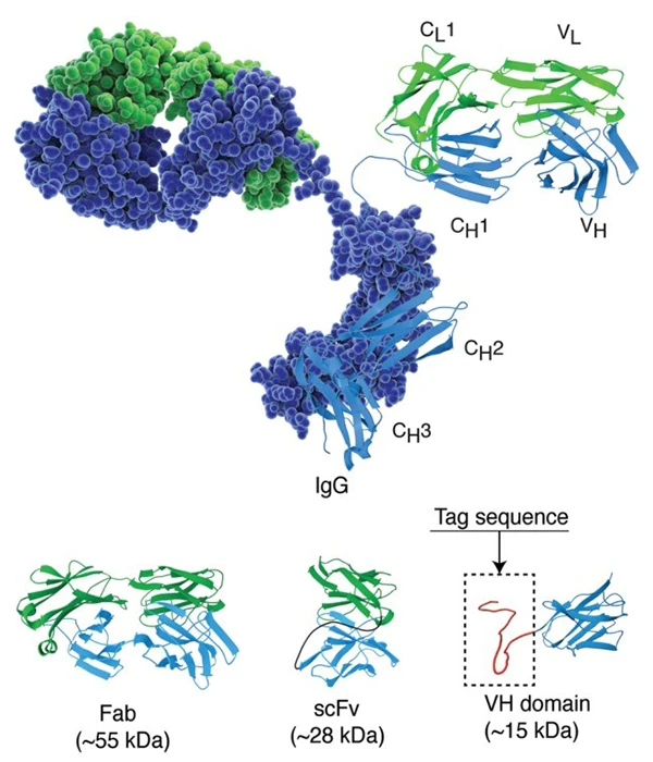
Image source: PMID: 25426869
The structure of immunoglobulin G in space-filling and cartoon representation with light and heavy chain colored in green and blue, respectively. Lower panel illustrates crystal structures of the antibody fragments FAB (fragment antigen-binding), scFv (single-chain fragment variable) and single domain antibody. A peptide tag of approximately 30 residues is illustrated at the c-terminal of the domain antibody. The immunoglobulin structure used in this figure is based on the RCSB Protein Data Bank entry 1igt.
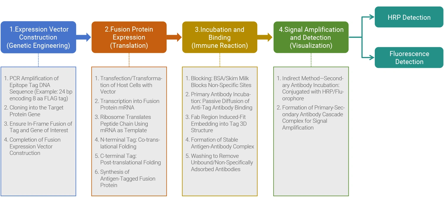
From Marker Protein Expression to Antibody Recognition, Usually divided intoFour key steps:
| Comparison Dimensions | Anti-tag antibody | Protein-specific antibody |
|---|---|---|
| Development Goals | For standardized tag sequences (such as FLAG, His) development | Unique epitopes specific to a particular natural protein |
| Development Cycle and Cost | Short development cycle (2-3 weeks), low cost, relies on hybridoma technology or recombinant expression platforms, with high batch-to-batch consistency. | Long development cycle (several months), high cost, and requires multiple rounds of validation for specificity. |
| Scope of Application | Any fusion protein (universal tool) bearing this tag | Recognize only specific natural proteins (specialized tool) |
| Detection Principle |
|
|
| Application Scenarios | Universal TestingPurification: Suitable for Western Blot, immunofluorescence, immunoprecipitation, affinity chromatography, etc. Supports high-throughput experiments and industrial production (e. g., antibody drug purification, enzyme preparation production, recombinant protein expression monitoring, protein interaction preliminary screening). | Specificity Studies: Suitable for scenarios such as protein-protein interaction network analysis, signaling pathway research, disease-related protein detection, and native conformation analysis, requiring differentiation between endogenous and exogenous proteins. |
| Core Advantages |
|
|
| Limitations |
|
|
Anti-tag antibodyAsA specific antibody targeting artificially introduced short peptide sequences, known as "tags," has become an extremely versatile and powerful tool in biomedical research. Its core advantage lies in the fact that researchers no longer need to develop and validate specific antibodies from scratch for each target protein. Instead, they can utilize existing, highly optimized anti-tag antibody systems to detect, purify, localize, and functionally manipulate a wide variety of proteins. The following sections will elaborate on its applications and core advantages.
In structural biology, obtaining high-quality protein crystals is a significant challenge. Anti-tag antibodies can serve as "crystallization chaperones" to facilitate the crystallization of target proteins. For example, the Fab fragment of the monoclonal antibody NZ-1, which recognizes the PA tag, can form a complex with the target protein containing the inserted PA tag, thereby promoting co-crystallization [1]. This method circumvents the difficulty of developing conformation-specific antibodies for each target protein by inserting a tag into specific loop regions of the target protein (such as β-hairpin structures). It leverages the high-affinity binding of antibodies to the tag to stabilize the protein conformation, ultimately yielding crystals suitable for high-resolution structural analysis.
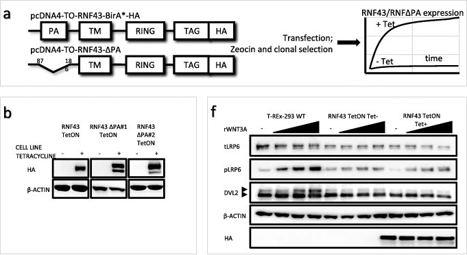
Image source: PMID: 30666745
Corresponding English translation:Inducible RNF43 and RNF43ΔPA expression in T-REx 293 cell line.
a. Schematic representation of the used experimental models. b. Western blot showing Tet-induced expression of HA-tagged RNF43 constructs in stable sell lines. f. Western blot analysis of canonical Wnt pathway activation by application of the increasing concentrations of rWNT3A (40, 60 and 100 ng/mL) after 3 h of treatment.
Chromatin immunoprecipitation is a key technique for studying protein-DNA interactions in vivo. When specific antibodies against endogenous transcription factors are unavailable or ineffective, expressing exogenous epitope-tagged fusion proteins and performing immunoprecipitation with highly specific anti-tag antibodies serves as a reliable and efficient alternative [2]. This method avoids the time-consuming and labor-intensive preparation of transgenic stable lines; for example, transient expression of tagged proteins in Arabidopsis protoplasts allows ChIP experiments to be completed within four days.
Anti-tag antibodies can be used for rapid, one-step affinity purification of recombinant proteins. For example, Sanae Tabata.Waiting for someone in 2010.Using the anti-TARGET tag antibody P20.1, a sensitive screening system can be established to rapidly select high-yield cell lines from mammalian cell culture supernatants and standardize the purification of milligram-scale target proteins. Similarly, in the production of Fv fragments, one-step affinity purification can be achieved through their fused Strep tag.
Anti-tag antibodies can not only "see" and "grab" but also "interfere with" and "activate" protein functions.
The anti-tag antibody system provides modular tools for constructing highly sensitive detection platforms.
In the clinical field, radionuclide-labeled anti-tag antibodies can be used for tumor localization and surgical navigation. For example, in recurrent colorectal cancer surgery, the use of iodine-125-labeled anti-tag antibodies (CC49 monoclonal antibodies) for radioimmunoguided surgery can detect more occult lesions that traditional imaging and surgical exploration cannot identify, thereby altering surgical plans and improving tumor resection rates.
Based on the above scenarios, the advantages of anti-tag antibodies are primarily reflected in the following aspects:
Traditional protein tags (such as GFP and HaloTag), while powerful, have relatively large molecular weights (typically over 20 kDa), which may interfere with the native structure, function, localization, and interactions of the protein of interest (POI). The new generation of ultra-small tag technologies, represented by the HiBiT peptide, is addressing these challenges.
The core advantages of the HiBiT tag lie in its extremely small size and its highly luminescent complementary system. HiBiT is a short peptide composed of only 11 amino acids (approximately 1.3 kDa). It does not emit light on its own but can bind with high affinity to a larger complementary protein fragment (LgBiT, 18 kDa) to form a complete and active NanoLuc luciferase. This design offers multiple revolutionary advantages:
The working principle of the nanobody-tag system involves camelid-derived or artificially designed single-domain antibodies that can bind with high affinity and specificity to short peptide sequences of 6–14 amino acids. These short peptide tags can be fused to the N-terminus or C-terminus of target proteins. After expression, the nanobodies serve as versatile tools for protein capture, visualization, or manipulation.
Although the interference from small tags and nanobodies has been minimized, in certain detailed studies, it is still necessary to completely remove the tags to obtain protein products that are entirely consistent with their native state. Cleavable tag systems have emerged to address this need, with the TEV protease cleavage site being one of the most commonly used and efficient systems.
TEV protease is a highly specific protease that recognizes its unique seven-amino-acid sequence (Glu-Asn-Leu-Tyr-Phe-Gln-Gly or similar variants) and cleaves between Gln and Gly/Ser. In protein purification or functional research workflows, the decision to introduce and ultimately remove tags is primarily based on the following considerations:
In bioengineering experiments, anti-tag antibodies are crucial tools that link target proteins with detection technologies. However, with the wide array of tag antibody products available on the market, how can one accurately select the right one based on experimental needs? We can From the experimental objective From tag and Tag Design to Antibody Characteristics and Technical Compatibility, Systematically Organize the Selection Strategy for Anti-Tag Antibodies to Help You Improve Experimental Efficiency and Data Reliability.
For protein localization studies, it is essential to visually present the subcellular distribution of target proteins through fluorescent signals or immunostaining. In such cases, fluorescent protein tags (such as GFP, mCherry) or small peptide tags (such as HA, FLAG) are more suitable.
Fluorescent tag: Directly provides visual signals without additional staining steps, suitable for live-cell imaging. The microscope excitation light source needs to be matched (e. g., GFP requires a 488 nm laser).
Small Peptide Tag: Compatible with fixed cell samples via immunofluorescence or immunohistochemistry detection, featuring a small tag size and minimal interference with protein function.
Protein purification is achieved by efficiently capturing the target protein through the high-affinity binding of a tag to a ligand, followed by its release under specific elution conditions, such as pH adjustment or the use of competitive agents. NeedSelect tags based on the characteristics of the target protein (e. g., prioritize His tags for membrane proteins, prioritize MBP tags for proteins prone to aggregation). AndAfter purification, the tag must be removed by enzymatic digestion or chemical cleavage to avoid interference with downstream functional experiments.
Common tags include His, GST, and MBP tags.
His-tag: Suitable for immobilized metal affinity chromatography (IMAC), with mild elution conditions (e. g., imidazole gradient), but attention should be paid to interference from endogenous histidine in host proteins.
GST tag: Eluted via glutathione, suitable for large-scale purification, but the tag is relatively large (26 kDa) may affect protein solubility.
MBP tag: Enhances solubility of target proteins, eluted with maltose, suitable for difficult-to-fold proteins.
Interaction studies require the capture of protein complexes through co-immunoprecipitation (Co-IP) or pull-down techniques, and the tags must meet the following criteria:
Small size: Avoids steric hindrance that could interfere with natural interactions (e. g., FLAG, HA, and Myc tags consist of only 8–10 amino acids).
High specificity: Antibodies must strictly recognize Do not tag sequences to avoid cross-reactions.
FLAG, HA, Myc tagsAvailableMinimize interference.
The molecular weight of the tag is recommended not to exceed 10% of the target protein (e. g., 50 kDa protein preferentially selects <5 kDa tag). A tag that is too large may interfere with the folding, stability, or interaction interface of the target protein. During the practical operation process,Predict the interface between the tag and the target protein using AlphaFold to avoid covering critical functional domains.
N-terminal tag: Suitable for cytoplasmic proteins without signal peptides, but may interfere with the transport of secreted proteins (e. g., N-terminal tag of antibody light chains can block secretion).
C-terminal tag: More versatile, but care must be taken to avoid disrupting localization signals at the C-terminus (such as mitochondrial targeting sequences).
Internal tag: Tags can be inserted into flexible loop regions of long proteins, requiring screening of mutation libraries to identify sites that do not affect function. Internal tag is suitable for functional key regions at the N/C-terminus of the target protein (such as the enzyme active center).
Monoclonal antibodies: High specificity, minimal batch-to-batch variation, suitable for quantitative experiments (e. g., WB, ELISA), but with high development costs.
Polyclonal antibodies: High sensitivity, capable of recognizing multiple epitopes, suitable for detecting low-abundance proteins, but require strict quality control for batch-to-batch variability.
For critical experiments (such as clinical sample testing), monoclonal antibodies are prioritized. Polyclonal antibodies can be used during preliminary experiments or screening stages to reduce costs.
Sensitivity: Select antibodies based on the expression level of the target protein (e. g., high-affinity antibodies are required for low-expression proteins, such as those with KD < 1 nM).
Specificity: Detect cross-reactivity of the target protein with host proteins (e. g., E.coli, HEK293) via Western blot (WB), prioritizing antibodies validated using knockout cell lines.
Validation Data: Verify that the antibody has been validated across multiple techniques such as WB, IP, IF, and ChIP, and avoid using antibodies validated solely by ELISA for IP experiments.
Primary antibody host: Commonly used hosts include rabbit, mouse, and goat. The primary antibody host species must match the secondary antibody host species (e. g., rabbit anti-FLAG primary antibody should be paired with anti-rabbit IgG secondary antibody). To avoid cross-binding of secondary antibodies, select primary antibodies from different host sources (e. g., mouse anti-HA + rabbit anti-Myc).
Formaldehyde Fixation: Cross-links proteins, potentially masking linear epitopes, necessitating the selection of antibodies that recognize conformational epitopes (such as certain fluorescently tag antibodies).
Methanol fixation: Suitable for membrane proteins, but may disrupt the antigenicity of certain tags (e. g., GST tags are prone to denaturation in methanol).
Heat-induced epitope retrieval (HIER): Suitable for formalin-fixed, paraffin-embedded sections, where epitopes are restored through high-temperature heating (e. g., 100°C for 20 minutes).
Enzyme Repair: Use Proteinase K or pepsin, optimizing concentration and time to avoid over-digestion.
Natural Proteins: Antibodies that recognize conformational epitopes should be selected (e. g., certain fluorescently tag antibodies only bind when the protein is correctly folded).
Denatured protein: Suitable for WB experiments, it is necessary to select antibodies that recognize linear epitopes (such as His-tag antibodies, which can still bind in SDS-PAGE).
For exampleInsert a FLAG tag at each end of the target protein (FLAG-protein-FLAG) to enhance detection signals via a dual-antibody sandwich assay. Tandem His6 tags (His6-His6) increase binding capacity to nickel columns, making them suitable for purifying low-expression proteins.
Purification + Detection Tags: For example, the His tag is used for purification, and the V5 tag is used for WB detection, to avoid interference from purification tags in downstream analysis.
Multicolor experimental tags: such as GFP (localization) + HA (interaction) + Myc (expression level), enabling multidimensional information acquisition.
CUSABIO offers high-quality products covering all scenarios. Rigorously validated across multiple scenarios.
Now available Free download complete Tag Antibody Product Rapid Selection, Facilitating Smooth Progress in Scientific Research!
Objective: Balance protein function, antibody accessibility, and structural integrity.
Operating Steps:
Objective: Minimize interference of tags on the conformation of the target protein and enhance antibody binding efficiency.
Operating Steps:
Objective: Select an expression system based on protein complexity, balancing yield with functional correctness.
Operating Steps:
Objective: Eliminate non-specific background signals.
Operating Steps:
Objective: Correct for antibody nonspecific binding (especially in IF and flow cytometry).
Operating Steps:
Objective: Verify the effectiveness of the experimental system.
Operating Steps:
| Fault Type | Possible reasons | Phenomenon Analysis | Solution |
|---|---|---|---|
| No signal or weak signal | Low expression level of the marker protein. | Low expression level of the marker protein. | Verification of Transfection/Transduction Efficiency: Use fluorescent co-transfection or flow cytometry to confirm the proportion of positive cells. |
| Optimization conditions: Conduct induction time/concentration gradient experiments and replace the promoter. | |||
| Check protein stability: Treat cells with proteasome inhibitors (such as MG132) and observe whether the signal is restored. | |||
| Insufficient Antibody Concentration | The positive control shows a weak signal, while the sample shows no signal. | Titration experiment: Incubate the same sample membrane with different dilutions (e. g., 1: 500, 1: 1000, 1: 5000) to determine the optimal concentration. | |
| Recommended starting dilution: For new antibodies, begin with the middle value of the range specified in the instructions (e. g., if the range is 1: 100-1000, use 1: 500). | |||
| Tag Epitope Masked | The tag antibody shows no signal, but the target protein-specific antibody shows a signal. | Protein Folding: Increase the sample boiling temperature (95°C, 10 min) or enhance the strength of the denaturant to fully expose the epitope. | |
| Alternative tag placement: If the N-terminal tag is blocked, attempt to construct a plasmid with a C-terminal tag. | |||
| Transfer Efficiency Issues | There is residual protein in the gel staining, or the markers on the membrane are unclear. | Protein size consideration: Large molecular proteins (>150 kDa) Choose wet transfer, low methanol, long time; small molecular weight proteins (<20 kDa) Use a 0.2 μm pore size membrane for a short duration. | |
| Transfer Optimization: After transfer, routinely perform Ponceau S staining to verify the transfer efficiency and observe loading consistency. | |||
| High Background Signal | Antibody Concentration Too High | The entire membrane appears blurred, with no clear bands visible. | Optimization Strategy: Reduce the concentration of the primary or secondary antibody, and shorten the incubation time. Pay special attention to the concentration of the secondary antibody, as it is often the main source of high background. |
| Inadequate Sealing | The background is uniform and has a matte texture. | Blocking Buffer Selection: Typically use 5% skim milk; switch to 5% BSA when detecting phosphorylated proteins (to avoid interference from casein in milk). | |
| Blocking Buffer Selection: Typically use 5% skim milk; switch to 5% BSA when detecting phosphorylated proteins (to avoid interference from casein in milk). | |||
| Inadequate Washing | The background shows localized patchy or streaky stains. | Wash Buffer Composition: TBST (containing Tween 20) is routinely used. | |
| Washing Duration and Frequency: Emphasize "small amounts, multiple times," with each session lasting 5-10 minutes, repeated more than 5 times. | |||
| Non-specific Binding | In addition to the target band, there is also a noticeable background of unintended bands. | Antibody Quality Issues: Use affinity-purified antibodies or monoclonal antibodies validated in the literature. | |
| Buffer Optimization: Increase the salt concentration or Tween-20 content in the antibody dilution/wash buffer to reduce hydrophobic interactions. | |||
| Multiple bands detected | Post-translational Modification | Bands larger than the target protein appear, or multiple bands with a regular pattern. | Expected modifications: Review literature to confirm known modifications. Perform de-modification treatments (e. g., dephosphorylation enzymes) as control verification. |
| Mobility Shift: Modifications such as phosphorylation/glycosylation can alter protein migration rates, which is a normal phenomenon. | |||
| Post-translational Modification | Bands larger than the target protein appear, or multiple bands with a regular pattern. | Expected modifications: Review literature to confirm known modifications. Perform de-modification treatments (e. g., dephosphorylation enzymes) as control verification. | |
| Mobility Shift: Modifications such as phosphorylation/glycosylation can alter protein migration rates, which is a normal phenomenon. | |||
| Variability in Myc Tag Recognition | There are many bands, even the internal reference channel has background bands. | Antibody Concentration Adjustment: Reduce the antibody concentration and perform a titration experiment. | |
| Additional Control: Set up knockdown (Knockdown) or knockout (Knockout) sample controls. If the band persists in the knockout sample, it is a non-specific band. | |||
| Tag-specific issues | HA tag is cleaved in apoptotic cells. | In addition to the full-length protein, there is an additional distinct small fragment. | Caspase cleavage site: The HA sequence contains a Caspase recognition site, which is cleaved during apoptosis. |
| Prevention Strategy: When conducting apoptosis experiments, switch to Flag or Myc tags; or simultaneously add tags at the C-terminus to verify the full-length protein. | |||
| Variability in Myc Tag Recognition | Western Blot can detect it, but immunofluorescence (IF) or immunoprecipitation (IP) cannot. | Fixed-dependent epitope accessibility: The Myc epitope is masked under specific fixatives (such as methanol) or in its native conformation. | |
| Solution: In IF experiments, try replacing the permeabilization agent (e. g., Triton X-100) or the fixative (e. g., 4% PFA). | |||
| Non-specific Binding of His-tag | The background is high during detection or purification, with many impurity proteins present. | Metal ion contamination: Host proteins containing histidine clusters can bind to nickel columns. | |
| Buffer composition adjustment: Add low-concentration imidazole (20-40 mM) to the binding/wash buffer to competitively elute contaminating proteins. |
[1] Tamura R, Miyazaki N, Ito Y, et al. Application of the NZ-1 Fab as a crystallization chaperone for PA tag-inserted target proteins. Protein Sci 2019; 28: 1450–59.
[2] Fang W, Wang X, Li Y, et al. Optimized protocols for chromatin immunoprecipitation of exogenously expressed epitope-tagged proteins. STAR Protoc 2023; 4: 102050.
[3] Hanl M, Li J, Schmidt T, et al. Target Engagement Studies and Kinetic Live-Cell Degradation Assays Enable the Systematic Characterization of Histone Deacetylase 6 Degraders. ACS Pharmacol Transl Sci 2025; 8: 2456–68.
[4] Boursier ME, Levin S, Zimmerman K, et al. The luminescent HiBiT peptide enables selective quantitation of G protein-coupled receptor ligand engagement and internalization in living cells. J Biol Chem 2020; 295: 5124–35.
[5] Yoda T, Shirai Y, Okada Y, et al. Four-color single-molecule imaging system for tracking GPCR dynamics with fluorescent HiBiT peptide. Biophys Physicobiol 2024; 21: e210020.
[6] Ankavay M, Dubuisson J, Cocquerel L, et al. Monitoring of hepatitis E virus infection and replication by functional tagging of the ORF2 protein. JHEP Rep 2025; 7: 101293.
[7] Cabalteja CC, Mihaylov D, Cubitt B, et al. Characterization of a Nanobody-Epitope Tag Interaction and Its Application for Receptor Engineering. ACS Chem Biol 2022; 17: 2345–56.
Antibody Reagent Types
Antibody Reagent Production
Antibody Reagent Applications
Further Reading