Call us
301-363-4651 (Available 9 a.m. to 5 p.m. CST from Monday to Friday)
| Code | CSB-BP015356MO |
| Abbreviation | Recombinant Mouse Myoc protein, partial |
| MSDS | |
| Size | $528 |
| Order now | |
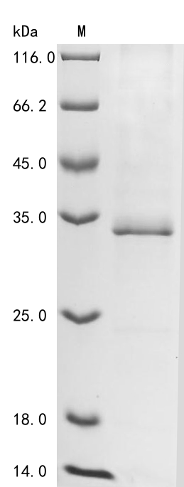
| Image |
|
| Have Questions? | Leave a Message or Start an on-line Chat |
Recombinant Mouse Myocilin (Myoc) is produced through a baculovirus expression system and covers amino acids 213-490—a partial but substantial portion of the protein. The recombinant protein includes an N-terminal 10xHis-tag and a C-terminal Myc-tag, which streamline purification and detection processes. SDS-PAGE analysis confirms purity levels exceeding 85%. This product is designed solely for research purposes, with no established biological functions or disease connections specified.
Myocilin appears to be a protein chiefly involved in extracellular matrix processes and can be found across various tissues, particularly in the eye. The protein likely plays an important role in maintaining structural integrity and supporting cellular signaling networks. Research into myocilin seems especially relevant for ocular health studies, given its apparent association with pathways that may influence intraocular pressure and other eye-related physiological processes.
Potential Applications
Note: The applications listed below are based on what we know about this protein's biological functions, published research, and experience from experts in the field. However, we haven't fully tested all of these applications ourselves yet. We'd recommend running some preliminary tests first to make sure they work for your specific research goals.
Mouse Myocilin is a complex extracellular glycoprotein that requires precise folding, proper oligomerization (trimer formation), specific tertiary structure, and glycosylation for its functional activity in ocular pressure regulation. The baculovirus expression system provides a eukaryotic environment that supports proper folding and potential glycosylation. However, the partial fragment (213-490aa) lacks critical N-terminal domains essential for full functionality and proper oligomerization. The dual N-terminal 10xHis-tag and C-terminal Myc-tag may cause significant steric interference with the protein's oligomerization interfaces and functional domains. While the expression system favors proper folding, the probability of correct folding with functional bioactivity is low due to the truncated nature and tag interference.
1. Protein-Protein Interaction Studies
This application carries a significant risk without functional validation. Myocilin interactions require proper oligomerization and native conformation. The partial fragment with dual tags may not form correct trimers and could exhibit non-specific binding or tag-mediated artefacts. If correctly folded and oligomerized (verified), it may identify physiological partners; if misfolded/unverified, interaction data will be biologically misleading.
2. Antibody Development and Validation
This application is highly suitable as antibody development relies on antigenic sequence recognition rather than functional protein folding. The fragment provides defined epitopes within the 213-490aa region for generating Myocilin-specific antibodies. The high purity ensures minimal contamination issues.
3. Biochemical Characterization Studies
These studies are essential for determining folding status but will not reflect native myocilin structure. Techniques should include size-exclusion chromatography to assess oligomeric state, circular dichroism spectroscopy to evaluate secondary structure, and glycosylation analysis. However, the partial fragment cannot form native trimers, and the results will describe an artificial construct.
Final Recommendation & Action Plan
The baculovirus-expressed Myocilin fragment with dual tags may be properly folded at the domain level, but cannot form functional oligomers due to the truncated nature and tag interference. Applications 2 (antibody development) can proceed immediately. Application 3 (biochemical characterization) provides limited insights into the fragment's properties but not native myocilin biology. Applications 1 should be avoided without rigorous validation of oligomerization capability. For reliable myocilin research, use a full-length protein expressed in mammalian systems that supports proper trimer formation and preserves native conformational epitopes.
There are currently no reviews for this product.

