TP53 Recombinant Monoclonal Antibody

Code CSB-RA236796A0HU
Size US$210
Order now
Image
  • Western Blot
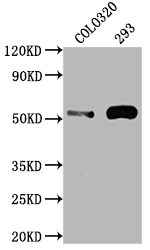
    Positive WB detected in: COLO320 whole cell lysate, 293 whole cell lysate
    All lanes: TP53 antibody at 1:2000
    Secondary
    Goat polyclonal to rabbit IgG at 1/50000 dilution
    Predicted band size: 44, 38, 39, 40, 34, 35, 30, 24, 25 kDa
    Observed band size: 53 kDa
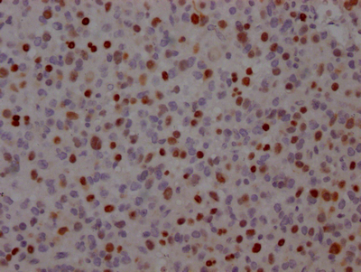
  • IHC image of CSB-RA236796A0HU diluted at 1:100 and staining in paraffin-embedded human glioma cancer performed on a Leica BondTM system. After dewaxing and hydration, antigen retrieval was mediated by high pressure in a citrate buffer (pH 6.0). Section was blocked with 10% normal goat serum 30min at RT. Then primary antibody (1% BSA) was incubated at 4℃ overnight. The primary is detected by a Goat anti-rabbit IgG polymer labeled by HRP and visualized using 0.05% DAB.
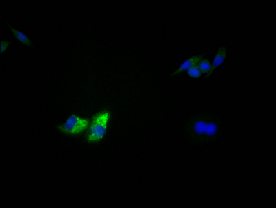
  • Immunofluorescence staining of HepG2 Cells with CSB-RA236796A0HU at 1:50, counter-stained with DAPI. The cells were fixed in 4% formaldehyde, permeated by 0.2% TritonX-100, and blocked in 10% normal Goat Serum. The cells were then incubated with the antibody overnight at 4℃. Nuclear DNA was labeled in blue with DAPI. The secondary antibody was FITC-conjugated AffiniPure Goat Anti-Rabbit IgG (H+L).
Have Questions? Leave a Message or Start an on-line Chat

Product Details

Uniprot No.
Target Names
Alternative Names
Cellular tumor antigen p53 (Antigen NY-CO-13) (Phosphoprotein p53) (Tumor suppressor p53), TP53, P53
Species Reactivity
Human
Immunogen
A synthesized peptide derived from human p53
Immunogen Species
Homo sapiens (Human)
Conjugate
Non-conjugated
Clonality
Monoclonal
Isotype
Rabbit IgG
Clone No.
3D4
Purification Method
Affinity-chromatography
Concentration
It differs from different batches. Please contact us to confirm it.
Buffer
Rabbit IgG in phosphate buffered saline, pH 7.4, 150mM NaCl, 0.02% sodium azide and 50% glycerol.
Form
Liquid
Tested Applications
ELISA, WB, IHC, IF
Recommended Dilution
Application Recommended Dilution
WB 1:500-1:5000
IHC 1:50-1:200
IF 1:20-1:200
Troubleshooting and FAQs
Storage
Upon receipt, store at -20°C or -80°C. Avoid repeated freeze.
Lead Time
Basically, we can dispatch the products out in 1-3 working days after receiving your orders. Delivery time maybe differs from different purchasing way or location, please kindly consult your local distributors for specific delivery time.
Description

The TP53 recombinant monoclonal antibody can detect human TP53 protein in ELISA, WB, IHC, and IF applications. It is produced using recombinant DNA technology, where the gene encoding the TP53 monoclonal antibody is synthesized after sequencing the cDNA of the TP53 antibody-producing hybridomas. To produce the hybridomas, B cells isolated from an animal immunized with a synthesized peptide derived from human TP53 are fused with myeloma cells. The synthesized gene is then cloned into a vector and transfected into cells for cultivation. The resulting TP53 recombinant monoclonal antibody is purified through affinity chromatography from the cell culture supernatant.

The TP53 protein, also known as p53, acts as a tumor suppressor by preventing cells from dividing when they have damaged DNA that could lead to the development of cancer. When DNA damage or other cellular stress occurs, the levels of p53 increase, which leads to the activation of multiple pathways that can result in several outcomes, including cell cycle arrest, apoptosis, senescence, and DNA repair. Mutations in TP53 that lead to loss of TP53 function can lead to uncontrolled cell growth and the development of tumors.

Customer Reviews and Q&A

 Customer Reviews

There are currently no reviews for this product.

Submit a Review here

Target Background

Function
Acts as a tumor suppressor in many tumor types; induces growth arrest or apoptosis depending on the physiological circumstances and cell type. Involved in cell cycle regulation as a trans-activator that acts to negatively regulate cell division by controlling a set of genes required for this process. One of the activated genes is an inhibitor of cyclin-dependent kinases. Apoptosis induction seems to be mediated either by stimulation of BAX and FAS antigen expression, or by repression of Bcl-2 expression. Its pro-apoptotic activity is activated via its interaction with PPP1R13B/ASPP1 or TP53BP2/ASPP2. However, this activity is inhibited when the interaction with PPP1R13B/ASPP1 or TP53BP2/ASPP2 is displaced by PPP1R13L/iASPP. In cooperation with mitochondrial PPIF is involved in activating oxidative stress-induced necrosis; the function is largely independent of transcription. Induces the transcription of long intergenic non-coding RNA p21 (lincRNA-p21) and lincRNA-Mkln1. LincRNA-p21 participates in TP53-dependent transcriptional repression leading to apoptosis and seems to have an effect on cell-cycle regulation. Implicated in Notch signaling cross-over. Prevents CDK7 kinase activity when associated to CAK complex in response to DNA damage, thus stopping cell cycle progression. Isoform 2 enhances the transactivation activity of isoform 1 from some but not all TP53-inducible promoters. Isoform 4 suppresses transactivation activity and impairs growth suppression mediated by isoform 1. Isoform 7 inhibits isoform 1-mediated apoptosis. Regulates the circadian clock by repressing CLOCK-ARNTL/BMAL1-mediated transcriptional activation of PER2.
Gene References into Functions
  1. Study summarizes the different functions of p53 in adipocyte development and in adipose tissue homeostasis. Furthermore, It explores the manipulation of p53 levels in adipose tissue depots and the impact on systemic energy metabolism in the context of insulin resistance and obesity. [review] PMID: 30181511
  2. a USP15-dependent lysosomal pathway controls p53-R175H turnover in ovarian cancer cells PMID: 29593334
  3. Results indicate that the underlying mechanisms whereby etoposide and ellipticine regulate CYP1A1 expression must be different and may not be linked to p53 activation alone. PMID: 29471073
  4. Studied association of tumor protein p53 and drug metabolizing enzyme polymorphisms with clinical outcome in patients with advanced nonsmall cell lung cancer. PMID: 28425245
  5. POH1 knockdown induced cell apoptosis through increased expression of p53 and Bim. PMID: 29573636
  6. a heretofore unappreciated effect of chronic high fat diet on beta-cells, wherein continued DNA damage owing to persistent oxidative stress results in p53 activation and a resultant inhibition of mRNA translation. PMID: 28630491
  7. diffuse large B cell lymphoma lacking CD19 or PAX5 expression were more likely to have mutant TP53. PMID: 28484276
  8. that proliferation potential-related protein promotes esophageal cancer cell proliferation and migration, and suppresses apoptosis by mediating the expression of p53 and IL-17 PMID: 30223275
  9. Infection of HIV-1 and subsequent HIV-1 reverse transcription are inhibited in HCT116 p53(+/+) cells in comparison to HCT116 p53(-/-) cells. Tumor suppressor gene p53 expression is upregulated in non-cycling cells. The restrictions of HIV by p53 is associated with the suppression of ribonucleotide reductase R2 subunit expression and phosphorylation of SAMHD1 protein. PMID: 29587790
  10. It has been shown that MDM2 and MDMX are targetable vulnerabilities within TP53-wild-type T-cell lymphomas. PMID: 29789628
  11. A significant increase in the expression of p53 and Bax was observed in cells treated with alpha-spinasterol, while cdk4/6 were significantly down-regulated upon exposure to alpha-spinasterol. PMID: 29143969
  12. There was a significant correlation between telomere dysfunction indices, p53, oxidative stress indices, and malignant stages of GI cancer patients PMID: 29730783
  13. PGEA-AN modulates P53 system which further leads to the death of the neuroblastoma cells with no effect on renal system in vivo owing it to be a future prospect for development of anticancer moiety against neuroblastoma. PMID: 29644528
  14. these data indicate that activation of autophagy reduces expression of STMN1 and p53, and the migration and invasion of cancer cells contributes to the anti-cancer effects of the Halofuginone. These findings may provide new insight into breast cancer prevention and therapy. PMID: 29231257
  15. miR-150 suppresses cigarette smoke-induced lung inflammation and airway epithelial cell apoptosis, which is causally linked to repression of p53 expression and NF-kappaB activity. PMID: 29205062
  16. tumors harboring TP53 mutations, which can impair epithelial function, have a unique bacterial consortium that is higher in relative abundance in smoking-associated tumors. PMID: 30143034
  17. crosstalk among p53, lipid metabolism, insulin resistance, inflammation and oxidative stress has roles in Non-alcoholic fatty liver disease [review] PMID: 30473026
  18. Ubiquitin-conjugating enzyme E2S (UBE2S) enhanced the ubiquitination of p53 protein to facilitate its degradation in hepatocellular carcinoma (HCC) cells. PMID: 29928880
  19. p53 knockout compensates osteopenia in murine Mysm1 deficiency. PMID: 29203593
  20. SIRT1 had a pivotally protective role in the regulation of ADSCs aging and apoptosis induced by H2O2 PMID: 29803744
  21. 133p53 promotes tumour invasion via IL-6 by activation of the JAK-STAT and RhoA-ROCK pathways. PMID: 29343721
  22. mutant TP53 G245C and R273H can lead to more aggressive phenotypes and enhance cancer cell malignancy. PMID: 30126368
  23. PD-L1, Ki-67, and p53 staining individually had significant prognostic value for patients with stage II and III colorectal cancer PMID: 28782638
  24. This study of patients with ccRCC, pooled analysis and multivariable modeling demonstrated that three recurrently mutated genes, BAP1, SETD2, and TP53, have statistically significant associations with poor clinical outcomes. Important clinical confounders, mutations of TP53 and SETD2 were associated with decreased CSS and RFS, respectively. PMID: 28753773
  25. Study revealed that the Wnt/beta-catenin signaling pathway and its major downstream target, c-Myc increased the miR552 levels and miR552 directly targets p53 tumor suppressor. miR552 may serve as an important link between functional loss of APC, leading to abnormal Wnt signals, and the absence of p53 protein in colorectal cancer. PMID: 30066856
  26. High levels of glucose leads to endothelial dysfunction via TAF1-mediated p53 Thr55 phosphorylation and subsequent GPX1 inactivation. PMID: 28673515
  27. Although tumor protein p53 (p53) does not directly control the luminal fate, its loss facilitates acquisition of mammary stem cell (MaSC)-like properties by luminal cells and predisposes them to development of mammary tumors with loss of luminal identity. PMID: 28194015
  28. Fifty-two percent of patients diagnosed with glioma/glioblastoma with a positive TP53 mutation. PMID: 29454261
  29. the expression of Ser216pCdc25C was also increased in the combined group, indicating that irinotecan likely radiosensitized the p53-mutant HT29 and SW620 cells through the ATM/Chk/Cdc25C/Cdc2 pathway. PMID: 30085332
  30. In the former, p53 binds to the CDH1 (encoding E-cadherin) locus to antagonize EZH2-mediated H3K27 trimethylation (H3K27me3) to maintain high levels of acetylation of H3K27 (H3K27ac). PMID: 29371630
  31. Among the hits, miR-596 was identified as a regulator of p53. The overexpression of miR-596 significantly increased p53 at the protein level, thereby inducing apoptosis. PMID: 28732184
  32. Apoptosis pathways are impaired in fibroblasts from patients with SSc, leading to chronic fibrosis. Nonetheless, PUMA/p53 pathway may not be involved in dysfunction of apoptosis mechanisms in fibroblasts of patients with SSc. PMID: 28905491
  33. Low TP53 expression is associated with drug resistance in colorectal cancer. PMID: 30106452
  34. The activation of p38 in response to low doses of ultraviolet radiation was postulated to be protective for p53-inactive cells. Therefore, MCPIP1 may favor the survival of p53-defective HaCaT cells by sustaining the activation of p38. PMID: 29103983
  35. TP53 missense mutations are associated with castration-resistant prostate cancer. PMID: 29302046
  36. P53 degradation is mediated by COP1 in the breast cancer. PMID: 29516369
  37. Combined inactivation of the XRCC4 non-homologous end-joining (NHEJ) DNA repair gene and p53 efficiently induces brain tumours with hallmark characteristics of human glioblastoma. PMID: 28094268
  38. A direct link between Y14 and p53 expression and suggests a function for Y14 in DNA damage signaling. PMID: 28361991
  39. TP53 Mutation is associated with Mouth Neoplasms. PMID: 30049200
  40. Cryo-Electron Microscopy studies on p53-bound RNA Polymerase II (Pol II) reveal that p53 structurally regulates Pol II to affect its DNA binding and elongation, providing new insights into p53-mediated transcriptional regulation. PMID: 28795863
  41. Increased nuclear p53 phosphorylation and PGC-1alpha protein content immediately following SIE but not CE suggests these may represent important early molecular events in the exercise-induced response to exercise PMID: 28281651
  42. E6/E7-p53-POU2F1-CTHRC1 axis promotes cervical cancer cell invasion and metastasis PMID: 28303973
  43. accumulated mutant-p53 protein suppresses the expression of SLC7A11, a component of the cystine/glutamate antiporter, system xC(-), through binding to the master antioxidant transcription factor NRF2. PMID: 28348409
  44. Consistently, forced expression of p53 significantly stimulated ACER2 transcription. Notably, p53-mediated autophagy and apoptosis were markedly enhanced by ACER2. Depletion of the essential autophagy gene ATG5 revealed that ACER2-induced autophagy facilitates its effect on apoptosis. PMID: 28294157
  45. results indicate that LGASC of the breast is a low-grade triple-negative breast cancer that harbours a basal-like phenotype with no androgen receptor expression, and shows a high rate of PIK3CA mutations but no TP53 mutations PMID: 29537649
  46. this study shows an inhibitory effect of wild-type P53 gene transfer on graft coronary artery disease in rat model PMID: 29425775
  47. Our results suggest that TP53 c.215G>C, p. (Arg72Pro) polymorphism may be considered as a genetic marker for predisposition to breast cancer in Moroccan population PMID: 29949804
  48. higher level of the p53 isoform, p53beta, predict better prognosis in patients with renal cell carcinoma through enhancing apoptosis in tumors. PMID: 29346503
  49. TP53 mutations are associated with colorectal liver metastases. PMID: 29937183
  50. High expression of TP53 is associated with oral epithelial dysplasia and oral squamous cell carcinoma. PMID: 29893337

Show More

Hide All

Involvement in disease
Esophageal cancer (ESCR); Li-Fraumeni syndrome (LFS); Squamous cell carcinoma of the head and neck (HNSCC); Lung cancer (LNCR); Papilloma of choroid plexus (CPP); Adrenocortical carcinoma (ADCC); Basal cell carcinoma 7 (BCC7)
Subcellular Location
Cytoplasm. Nucleus. Nucleus, PML body. Endoplasmic reticulum. Mitochondrion matrix. Cytoplasm, cytoskeleton, microtubule organizing center, centrosome.; [Isoform 1]: Nucleus. Cytoplasm. Note=Predominantly nuclear but localizes to the cytoplasm when expressed with isoform 4.; [Isoform 2]: Nucleus. Cytoplasm. Note=Localized mainly in the nucleus with minor staining in the cytoplasm.; [Isoform 3]: Nucleus. Cytoplasm. Note=Localized in the nucleus in most cells but found in the cytoplasm in some cells.; [Isoform 4]: Nucleus. Cytoplasm. Note=Predominantly nuclear but translocates to the cytoplasm following cell stress.; [Isoform 7]: Nucleus. Cytoplasm. Note=Localized mainly in the nucleus with minor staining in the cytoplasm.; [Isoform 8]: Nucleus. Cytoplasm. Note=Localized in both nucleus and cytoplasm in most cells. In some cells, forms foci in the nucleus that are different from nucleoli.; [Isoform 9]: Cytoplasm.
Protein Families
P53 family
Tissue Specificity
Ubiquitous. Isoforms are expressed in a wide range of normal tissues but in a tissue-dependent manner. Isoform 2 is expressed in most normal tissues but is not detected in brain, lung, prostate, muscle, fetal brain, spinal cord and fetal liver. Isoform 3
Database Links

HGNC: 11998

OMIM: 133239

KEGG: hsa:7157

STRING: 9606.ENSP00000269305

UniGene: Hs.437460

CUSABIO guaranteed quality
icon of phone
Call us
301-363-4651 (Available 9 a.m. to 5 p.m. CST from Monday to Friday)
icon of address
Address
7505 Fannin St., Ste 610, Room 7 (CUBIO Innovation Center), Houston, TX 77054, USA
icon of social media
Join us with

Subscribe newsletter

Leave a message

* To protect against spam, please pass the CAPTCHA test below.
CAPTCHA verification
© 2007-2024 CUSABIO TECHNOLOGY LLC All rights reserved. 鄂ICP备15011166号-1
Place an order now

I. Product details

*
*
*
*

II. Contact details

*
*

III. Ship To

*
*
*
*
*
*
*

IV. Bill To

*
*
*
*
*
*
*
*