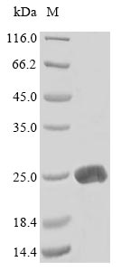
The process of recombinant mouse Lymphotoxin-alpha (Lta) production starts with the identification and isolation of the gene that codes for the Lta protein (34-202aa). This gene is cloned into a plasmid vector along with the N-terminal 10xHis-tag and C-terminal Myc-tag gene, which is introduced into E. coli cells. The E. coli cells are cultured in bioreactors to express the protein. Once the cells have grown sufficiently, they are lysed to release the protein. The Lta protein is purified using affinity chromatography, with a purity of up to 85% as determined by SDS-PAGE.
Lta a cytokine within the TNF superfamily [1], plays a pivotal role in the development of lymphoid organs, cellular cytotoxicity, and immune system regulation [2]. Lta pairs with lymphotoxin-beta (LT-β) to form a heteromeric complex, integral to various immunological functions [3]. Research indicates that Lta is essential for forming lymphoid tissues, including germinal centers, and is critical for lymphoid organogenesis [4].
Lta is also associated with pro-inflammatory responses, enhancing T-cell activity and influencing survival rates in conditions such as melanoma [5]. Furthermore, Lta is vital for the development of intestinal lymphoid organs, like Peyer's patches and mesenteric lymph nodes [6]. In systemic lupus erythematosus, B cells secrete Lta and other pro-inflammatory cytokines, driving the inflammatory process and promoting the formation of germinal centers in inflamed tissues [7].
References:
[1] B. Paik and L. Tong, Polymorphisms in lymphotoxin-alpha as the “missing link” in prognosticating favourable response to omega-3 supplementation for dry eye disease: a narrative review, International Journal of Molecular Sciences, vol. 24, no. 4, p. 4236, 2023. https://doi.org/10.3390/ijms24044236
[2] R. Bates, E. Browne, R. Schalks, H. Jacobs, L. Tan, P. Parekhet al., Lymphotoxin-alpha expression in the meninges causes lymphoid tissue formation and neurodegeneration, Brain, vol. 145, no. 12, p. 4287-4307, 2022. https://doi.org/10.1093/brain/awac232
[3] P. Lawton, J. Nelson, R. Tizard, & J. Browning, Characterization of the mouse lymphotoxin-beta gene., The Journal of Immunology, vol. 154, no. 1, p. 239-246, 1995. https://doi.org/10.4049/jimmunol.154.1.239
[4] P. Koni, R. Sacca, P. Lawton, J. Browning, N. Ruddle, & R. Flavell, Distinct roles in lymphoid organogenesis for lymphotoxins α and β revealed in lymphotoxin β–deficient mice, Immunity, vol. 6, no. 4, p. 491-500, 1997. https://doi.org/10.1016/s1074-7613(00)80292-7
[5] S. Yang, K. Hayer, H. Fazelinia, L. Spruce, M. Asnani, A. Naqviet al., Fbxw7β isoform drives transcriptional activation of the proinflammatory tnf cluster in human pro-b cells, Blood Advances, vol. 7, no. 7, p. 1077-1091, 2023. https://doi.org/10.1182/bloodadvances.2022007910
[6] T. Spahn, M. Müller, W. Domschke, & T. Kucharzik, Role of lymphotoxins in the development of peyer's patches and mesenteric lymph nodes, Annals of the New York Academy of Sciences, vol. 1072, no. 1, p. 187-193, 2006. https://doi.org/10.1196/annals.1326.029
[7] Y. Yusof, E. Vital, & P. Emery, B-cell-targeted therapies in systemic lupus erythematosus and anca-associated vasculitis: current progress, Expert Review of Clinical Immunology, vol. 9, no. 8, p. 761-772, 2013. https://doi.org/10.1586/1744666x.2013.816479


