Call us
301-363-4651 (Available 9 a.m. to 5 p.m. CST from Monday to Friday)
| Code | CSB-CF3574GMC |
| Abbreviation | Recombinant Influenza A virus M2 protein |
| MSDS | |
| Size | $1620 |
| Order now | |
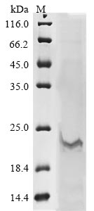
| Image |
|
| Have Questions? | Leave a Message or Start an on-line Chat |
Recombinant Influenza A virus Matrix protein 2 (M2) is produced through an in vitro E.coli expression system, covering the complete 1-97 amino acid sequence. The protein comes with an N-terminal 10xHis-tag and a C-terminal Myc-tag, which makes purification and detection more straightforward. SDS-PAGE analysis shows purity levels exceeding 85%. This research-use-only product works well for experimental applications that need high-quality viral proteins.
The Matrix protein 2 (M2) of Influenza A virus appears to play a critical role in the virus's life cycle, especially during viral uncoating and assembly. It works as an ion channel, which seems vital for acidifying the viral interior—a process necessary for genome release. M2 has become a key target in antiviral research and may provide valuable insights into how influenza virus replication and pathogenesis actually work.
Potential Applications
Note: The applications listed below are based on what we know about this protein's biological functions, published research, and experience from experts in the field. However, we haven't fully tested all of these applications ourselves yet. We'd recommend running some preliminary tests first to make sure they work for your specific research goals.
Based on the provided information, the recombinant Influenza A virus M2 protein is unlikely to be correctly folded or bioactive. M2 is a transmembrane protein that requires proper insertion into a lipid bilayer for correct folding and oligomerization into a functional tetrameric proton channel. The cell-free E. coli expression system lacks a membrane environment and eukaryotic chaperones, which are essential for transmembrane protein folding. The dual tags (N-terminal 10xHis and C-terminal Myc) may further disrupt the protein's structure, as the tags could interfere with the transmembrane domain or oligomerization sites. The purity of >85% indicates some impurities, but does not guarantee proper folding. Without experimental validation (e.g., circular dichroism for secondary structure, proton channel activity assays, or oligomerization checks), the protein cannot be assumed to be functional.
1. Antibody Development and Characterization
This recombinant M2 protein can be used for antibody development, but if misfolded, antibodies may primarily recognize linear epitopes or tag-derived sequences rather than conformational epitopes of native M2. The dual tags may dominate the immune response, reducing specificity for viral M2. If correctly folded, antibodies might recognize native M2; however, without folding validation, antibodies should be validated against full-length, properly folded M2 or viral particles to ensure relevance. The full-length sequence is advantageous but does not guarantee correct conformation.
2. Protein-Protein Interaction Studies
The dual-tagged M2 protein can be employed in pull-down assays, but if misfolded, interactions may be tag-mediated or non-specific, leading to false positives. The tags allow immobilization, but biologically relevant binding partners (e.g., host factors during infection) may not bind without native conformation. If used, validate folding first and include controls (e.g., tag-only proteins or inactive mutants) to distinguish specific interactions. Results should be confirmed with eukaryotic-expressed M2.
3. Biochemical and Structural Analysis
This recombinant M2 is unsuitable for meaningful structural analysis without folding validation and tag removal. The tags may alter biophysical properties (e.g., in circular dichroism or dynamic light scattering), and the lack of a membrane environment means data may not reflect native structure. If misfolded, stability or oligomerization studies will be invalid. For reliable insights, use membrane-reconstituted M2 and remove tags for high-resolution techniques like crystallography.
4. Assay Development and Screening Applications
The dual-tagged M2 can be used as a standard in assays (e.g., ELISA or Western blot), but the tags may affect antibody binding, leading to inaccurate quantification. If misfolded, it may not serve as a reliable positive control for native M2 detection. For screening applications (e.g., small molecule interactions), validate that the protein's conformation mimics native M2. Use with caution and include controls to account for tag interference.
Final Recommendation & Action Plan
To ensure reliable results, first validate the folding and bioactivity of the recombinant M2 protein using techniques such as circular dichroism to assess secondary structure, size-exclusion chromatography or blue native PAGE to check oligomerization, and proton flux assays to confirm channel activity. Given the challenges of expressing transmembrane proteins in cell-free systems, consider using eukaryotic expression (e.g., mammalian cells) for functional studies. If using this protein, remove the tags via proteolytic cleavage and reconstitute the protein in liposomes for biochemical assays. For antibody development, validate antibodies with native M2 from viral particles. In all applications, include appropriate controls, such as tag-free proteins and known inhibitors, to minimize artifacts. Prioritize folding validation before any functional or immunological studies.
There are currently no reviews for this product.

