Call us
301-363-4651 (Available 9 a.m. to 5 p.m. CST from Monday to Friday)
| Code | CSB-RA587302A0HU |
| Size | US$210 |
| Order now | |
| Image |
|
| Have Questions? | Leave a Message or Start an on-line Chat |
| Application | Recommended Dilution |
|---|---|
| WB | 1:500-1:5000 |
ITCH is an E3 ubiquitin-protein ligase that plays a central role in regulating immune responses, cell signaling pathways, and protein turnover through targeted ubiquitination of substrate proteins. As a HECT-type ubiquitin ligase, ITCH modulates the stability and activity of key signaling molecules, making it an important target for researchers investigating ubiquitin-mediated proteolysis, inflammatory signaling, and cellular homeostasis.
This recombinant monoclonal antibody, clone 9C3, offers the consistency and reliability that demanding experimental workflows require. Because it is produced from a defined genetic sequence rather than traditional hybridoma methods, you can expect uniform performance across lots, eliminating the variability that can complicate long-term studies or multi-site collaborations. The rabbit IgG format provides excellent signal-to-noise characteristics, while affinity chromatography purification ensures high specificity for your target.
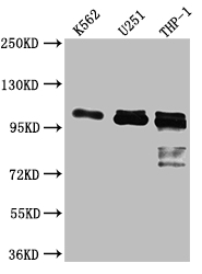
Validation studies confirm robust performance in Western blot applications, where the antibody successfully detects ITCH in human cell lysates from K562, U-251, and THP-1 cells at dilutions ranging from 1:500 to 1:5000. The observed band at 103 kDa aligns with the predicted molecular weight, providing confidence in target specificity. The diverse cell line panel spanning hematopoietic and neural lineages demonstrates reliable detection across different cellular contexts. ELISA compatibility extends your experimental options for quantitative analyses.
Whether you are investigating ubiquitin pathway dynamics, characterizing protein degradation mechanisms, or exploring ITCH's role in immune cell function, this antibody delivers the reproducible results needed to advance your cell biology research with confidence.
Applications : Immunohistochemistry (IHC)
Sample type: cell
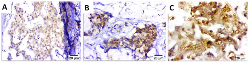
Review: Representative photomicrographs for immunohistochemical staining of ITCH. Invasive ductal carcinoma showing moderate [A] and strong [B] cytoplasmic and membranous staining of CXCR4. Brown color indicates positive staining. Negative, weak, moderate and strong staining = 0, 1, 2 and 3, respectively. Magnification: ×400, scale bar: 20 μm.
By Anonymous

