Recombinant Sudan ebolavirus Envelope glycoprotein (GP), partial

In Stock
Code CSB-EP742487SRE
Abbreviation Recombinant Sudan ebolavirus GP protein, partial
MSDS
Size $388
Order now
Image
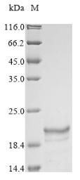
  • (Tris-Glycine gel) Discontinuous SDS-PAGE (reduced) with 5% enrichment gel and 15% separation gel.
Have Questions? Leave a Message or Start an on-line Chat

Product Details

Purity
Greater than 85% as determined by SDS-PAGE.
Target Names
GP
Uniprot No.
Research Area
Others
Alternative Names
(GP1,2)(GP)
Species
Sudan ebolavirus (strain Uganda-00) (SEBOV) (Sudan Ebola virus)
Source
E.coli
Expression Region
502-637aa
Target Protein Sequence
QTNTKATGKCNPNLHYWTAQEQHNAAGIAWIPYFGPGAEGIYTEGLMHNQNALVCGLRQLANETTQALQLFLRATTELRTYTILNRKAIDFLLRRWGGTCRILGPDCCIEPHDWTKNITDKINQIIHDFIDNPLPN
Note: The complete sequence may include tag sequence, target protein sequence, linker sequence and extra sequence that is translated with the protein sequence for the purpose(s) of secretion, stability, solubility, etc.
If the exact amino acid sequence of this recombinant protein is critical to your application, please explicitly request the full and complete sequence of this protein before ordering.
Mol. Weight
30.8 kDa
Protein Length
Partial
Tag Info
N-terminal 6xHis-KSI-tagged
Form
Liquid or Lyophilized powder
Note: We will preferentially ship the format that we have in stock, however, if you have any special requirement for the format, please remark your requirement when placing the order, we will prepare according to your demand.
Buffer
If the delivery form is liquid, the default storage buffer is Tris/PBS-based buffer, 5%-50% glycerol. If the delivery form is lyophilized powder, the buffer before lyophilization is Tris/PBS-based buffer, 6% Trehalose.
Reconstitution
We recommend that this vial be briefly centrifuged prior to opening to bring the contents to the bottom. Please reconstitute protein in deionized sterile water to a concentration of 0.1-1.0 mg/mL.We recommend to add 5-50% of glycerol (final concentration) and aliquot for long-term storage at -20°C/-80°C. Our default final concentration of glycerol is 50%. Customers could use it as reference.
Troubleshooting and FAQs
Storage Condition
Store at -20°C/-80°C upon receipt, aliquoting is necessary for mutiple use. Avoid repeated freeze-thaw cycles.
Shelf Life
The shelf life is related to many factors, storage state, buffer ingredients, storage temperature and the stability of the protein itself.
Generally, the shelf life of liquid form is 6 months at -20°C/-80°C. The shelf life of lyophilized form is 12 months at -20°C/-80°C.
Lead Time
3-7 business days
Notes
Repeated freezing and thawing is not recommended. Store working aliquots at 4°C for up to one week.
Datasheet & COA
Please contact us to get it.
Description

Recombinant Sudan ebolavirus Envelope glycoprotein (GP) is produced in E. coli, covering amino acid region 502-637. This partial protein carries an N-terminal 6xHis-KSI tag to help with purification and achieves over 85% purity as confirmed by SDS-PAGE. The preparation appears suitable for various research applications and should provide reliable, reproducible results.

The Sudan ebolavirus Envelope glycoprotein (GP) likely plays a crucial role in the viral lifecycle, particularly in mediating entry into host cells. It seems essential for fusion of the viral membrane with the host cell membrane, which makes it a significant focus in virology research. Understanding its structure and interactions may be vital for developing therapeutic approaches against ebolavirus infections.

Potential Applications

Note: The applications listed below are based on what we know about this protein's biological functions, published research, and experience from experts in the field. However, we haven't fully tested all of these applications ourselves yet. We'd recommend running some preliminary tests first to make sure they work for your specific research goals.

1. Antibody Development and Epitope Mapping

This recombinant GP fragment (502-637aa) could serve as an immunogen for generating polyclonal or monoclonal antibodies specific to the Sudan ebolavirus envelope glycoprotein. The N-terminal 6xHis-KSI tag helps with purification and immobilization for immunization protocols. The fragment represents a specific region of the GP that might contain unique epitopes distinct from other ebolavirus strains. Researchers could potentially use this protein to develop strain-specific antibodies for research applications and comparative studies between different ebolavirus species.

2. Protein-Protein Interaction Studies

The 6xHis-KSI tagged GP fragment can be used in pull-down assays to identify potential cellular binding partners or co-factors that interact with this specific region of the Sudan ebolavirus envelope glycoprotein. The histidine tag allows efficient immobilization on nickel-based resins for affinity purification experiments. Researchers can incubate cell lysates or purified protein libraries with the immobilized GP fragment to capture interacting proteins for subsequent mass spectrometry analysis. This approach might reveal host cell factors involved in viral entry or membrane fusion processes.

3. ELISA-Based Binding Assays

The recombinant GP fragment with its N-terminal tag can be used as a coating antigen in enzyme-linked immunosorbent assays to study binding interactions with various ligands, receptors, or other proteins. The 85% purity level appears sufficient for ELISA applications where the protein can be directly coated onto microplate wells. Researchers can investigate binding specificity, affinity measurements, and competitive binding studies using this fragment as a capture reagent. The tag also permits oriented immobilization using anti-His antibodies or nickel-coated plates.

4. Biochemical Characterization and Stability Studies

This partial GP protein can be subjected to various biochemical analyses to characterize its biophysical properties, including thermal stability, pH sensitivity, and aggregation behavior. The fragment's stability under different buffer conditions, temperature ranges, and storage conditions can be monitored using techniques such as dynamic light scattering, circular dichroism spectroscopy, or analytical ultracentrifugation. Such studies may provide valuable insights into the structural properties of this specific GP region and inform optimal conditions for experimental use and storage.

Customer Reviews and Q&A

 Customer Reviews

There are currently no reviews for this product.

Submit a Review here

Target Background

Function
Trimeric GP1,2 complexes form the virion surface spikes and mediate the viral entry processes, with GP1 acting as the receptor-binding subunit and GP2 as the membrane fusion subunit. At later times of infection, downregulates the expression of various host cell surface molecules that are essential for immune surveillance and cell adhesion. Down-modulates several integrins including ITGA1, ITGA2, ITGA3, ITGA4, ITGA5, ITGA6, ITGAV and ITGB1. This decrease in cell adhesion molecules may lead to cell detachment, contributing to the disruption of blood vessel integrity and hemorrhages developed during infection (cytotoxicity). Interacts with host TLR4 and thereby stimulates the differentiation and activation of monocytes leading to bystander death of T-lymphocytes. Downregulates as well the function of host natural killer cells. Counteracts the antiviral effect of host BST2/tetherin that restricts release of progeny virions from infected cells. However, cooperates with VP40 and host BST2 to activate canonical NF-kappa-B pathway in a manner dependent on neddylation.; Functions as a decoy for anti-GP1,2 antibodies thereby contributing to viral immune evasion. Interacts and activates host macrophages and dendritic cells inducing up-regulation of cytokine transcription. This effect is mediated throught activation of host TLR4.; Responsible for binding to the receptor(s) on target cells. Interacts with CD209/DC-SIGN and CLEC4M/DC-SIGNR which act as cofactors for virus entry into dendritic cells (DCs) and endothelial cells. Binding to the macrophage specific lectin CLEC10A also seems to enhance virus infectivity. Interaction with FOLR1/folate receptor alpha may be a cofactor for virus entry in some cell types, although results are contradictory. Members of the Tyro3 receptor tyrosine kinase family also seem to be cell entry factors in filovirus infection. Once attached, the virions are internalized through clathrin-dependent endocytosis and/or macropinocytosis. After internalization of the virus into the endosomes of the host cell, proteolysis of GP1 by two cysteine proteases, CTSB/cathepsin B and CTSL/cathepsin L removes the glycan cap and allows GP1 binding to the host entry receptor NPC1. NPC1-binding, Ca(2+) and acidic pH induce a conformational change of GP2, which unmasks its fusion peptide and permit membranes fusion.; Acts as a class I viral fusion protein. Under the current model, the protein has at least 3 conformational states: pre-fusion native state, pre-hairpin intermediate state, and post-fusion hairpin state. During viral and target cell membrane fusion, the coiled coil regions (heptad repeats) assume a trimer-of-hairpins structure, positioning the fusion peptide in close proximity to the C-terminal region of the ectodomain. The formation of this structure appears to drive apposition and subsequent fusion of viral and target cell membranes. Responsible for penetration of the virus into the cell cytoplasm by mediating the fusion of the membrane of the endocytosed virus particle with the endosomal membrane. Low pH in endosomes induces an irreversible conformational change in GP2, releasing the fusion hydrophobic peptide.
Subcellular Location
[GP2]: Virion membrane; Single-pass type I membrane protein. Host cell membrane; Single-pass type I membrane protein.; [GP1]: Virion membrane; Peripheral membrane protein. Host cell membrane; Peripheral membrane protein.; [Shed GP]: Secreted.
Protein Families
Filoviruses glycoprotein family
Database Links

KEGG: vg:3160774

CUSABIO guaranteed quality
icon of phone
Call us
301-363-4651 (Available 9 a.m. to 5 p.m. CST from Monday to Friday)
icon of address
Address
7505 Fannin St., Ste 610, Room 7 (CUBIO Innovation Center), Houston, TX 77054, USA
icon of social media
Join us with

Subscribe newsletter

Leave a message

* To protect against spam, please pass the CAPTCHA test below.
CAPTCHA verification
© 2007-2025 CUSABIO TECHNOLOGY LLC All rights reserved. 鄂ICP备15011166号-1
Place an order now

I. Product details

*
*
*
*

II. Contact details

*
*

III. Ship To

*
*
*
*
*
*
*

IV. Bill To

*
*
*
*
*
*
*
*