Call us
301-363-4651 (Available 9 a.m. to 5 p.m. CST from Monday to Friday)
Kidney cells, as implied in its name, refer to the cells in kidney. Before knowing about kidney cells, we learn some knowledge of kidney. The kidney is an organ that is responsible for filtering the blood and making urine. The kidney is a powerful chemical factory that performs the critical regulation of the body's salt, potassium and acid content. Everybody has two kidneys that are located on either side of the spine at the lowest level of the rib cage with the size of a fist. Each kidney contains up to a million functional units named nephrons. A nephron consists of a filtering unit of tiny blood vessels called a glomerulus, which is attached to a tubule. When blood enters the glomerulus, it is filtered and the remaining fluid then passes along the tubule. In the tubule, chemicals and water are either added to or removed from this filtered fluid according to the body's demands, the final product is the urine excreted by us. Cells are the basic unit of organ formation and function. So what are the types of cells in kidney? Let’s start to know more about the types of kidney cells.
There are many different cell types in the kidney, usually including tubule epithelial cell, macula densa cell, glomerular endothelial cell, podocyte, mesangial cell and parietal epithelial cell (Figure 1).
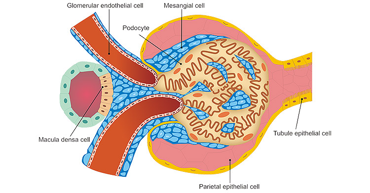
Figure 1. The common types of kidney cells
Tubule epithelial cell refers to the outer layer of cells of the renal tubules, which is able to reabsorb all the glucose and amino acids in the glomerular filtrate and excrete other non-nutrients into the urine. It plays a critical role in renal function. Tubular epithelial cells is the main site of injury in metabolic and inflammatory diseases. These cells can secrete several inflammatory mediators including cytokines and chemokines, and actively participate in the acute inflammatory process by producing IL-8 to stimulate the differentiation of leukocytes.
Podocyte is terminally differentiated cell of the kidney glomerulus that is able to promote the development of glomeruli, resist intraglomerular pressure, maintain vascular loop shape and regulate glomerular filtration rate. Moreover, this cell also can produce VEGF to regulate endothelial cells, participate in inflammation and immune response, synthesize and decompose glomerular basement membrane. Dysfunction of glomerular podocytes and subsequent cellular death were found to be the driving forces behind disease initiation and progression, respectively [1].
Parietal epithelial cell (PEC) and visceral podocyte make up the epithelial cells of the renal glomerulus. PEC lines the inner surface of Bowman’s capsule. A large evidence has recently suggested that PEC represents a reservoir of renal progenitors in adult human kidney which generate novel podocytes during childhood and adolescence, and can regenerate injured podocytes [2]. These evidence suggests that podocyte injury can be repaired.
Mesangial cell is a specialized pericyte that surrounds and constrains the vascular network within the glomerulus of the kidney. These cells are derived from the stromal mesenchyme, a progenitor population distinct from nephron stem cells [3]. It has a variety of functlons, including synthesis and assembly of the mesangial matrix, endocytosis and processing of plasma macro-molecules, and control of glomerular hemodynamics via mesangial cell contraction or release of vasoactive hormones.
Macula densa cell is renal sensor element that detect changes in distal tubular fluid composition and transmit signals to the glomerular vascular elements. This tubuloglomerular feedback mechanism plays an important role in regulating glomerular filtration rate and blood flow. Macula densa cell detects changes in luminal sodium chloride concentration through a complex series of ion transport-related intracellular events [4].
Cell Markers refer to a series of proteins, which can distinguish the special type of cells from other types. In this part, we list partial secreted factors and cell markers of these types of kidney cells.
References:
[1] Reiser J, Sever S. Podocyte biology and pathogenesis of kidney disease [J]. Annu Rev Med. 2013;64:357-366.
[2] Romagnani P. Parietal epithelial cells: their role in health and disease [J]. Contrib Nephrol. 2011;169:23-36.
[3] Boyle SC, Liu Z, Kopan R. Notch signaling is required for the formation of mesangial cells from a stromal mesenchyme precursor during kidney development [J]. Development. 2014;141(2):346-354.
[4] Bell PD, Lapointe JY, Peti-Peterdi J. Macula densa cell signaling [J]. Annu Rev Physiol. 2003;65:481-500.
Categories of Cell Marker
Cell Marker Related Articles
Cell Marker Related Pathways