Call us
301-363-4651 (Available 9 a.m. to 5 p.m. CST from Monday to Friday)
| Code | CSB-EP326185BKF |
| Abbreviation | Recombinant Bovine leukemia virus env protein, partial |
| MSDS | |
| Size | $9.9 |
| Promotion |

|
| Order now | |
| Image |
|
| Have Questions? | Leave a Message or Start an on-line Chat |
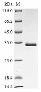
This recombinant Bovine leukemia virus Envelope glycoprotein (env) comes from E. coli expression and spans amino acids 34 to 301. The protein carries an N-terminal 10xHis-tag and a C-terminal Myc-tag, which should make purification and detection more straightforward. SDS-PAGE analysis shows purity levels above 90%, suggesting it may perform reliably in research settings. What researchers get is a partial sequence that appears suitable for different experimental approaches.
The Envelope glycoprotein of Bovine leukemia virus seems critical for viral entry—it's what likely handles the first contact with host cells. Being part of the viral envelope, this protein probably plays an important role during infection. For researchers studying viral mechanisms and host-pathogen interactions, understanding this protein could be valuable. Its significance in research comes from what it might reveal about viral spread and possible ways to intervene.
Potential Applications
Note: The applications listed below are based on what we know about this protein's biological functions, published research, and experience from experts in the field. However, we haven't fully tested all of these applications ourselves yet. We'd recommend running some preliminary tests first to make sure they work for your specific research goals.
Bovine Leukemia Virus (BLV) envelope glycoprotein is a viral surface protein that almost certainly requires extensive glycosylation and complex eukaryotic folding machinery (including disulfide bond formation) for correct tertiary structure and bioactivity. E. coli, a prokaryotic system, cannot perform glycosylation and often fails to properly fold complex eukaryotic viral glycoproteins. The protein is also a partial fragment (34-301aa), which may lack critical structural domains. Therefore, while not certain without experimental data, it is probable that this recombinant protein is misfolded, unglycosylated, and lacks the native bioactivity (e.g., receptor binding) of the viral envelope protein.
1. Antibody Development and Characterization
This application is feasible, but with a major caveat. The recombinant envelope glycoprotein can be used as an immunogen to generate antibodies. However, because the protein is likely unglycosylated and misfolded, the resulting antibodies will primarily recognize linear epitopes of the denatured protein backbone. They may have poor or no reactivity against the native, glycosylated form of the BLV envelope glycoprotein present on actual virions or in infected cells. Antibody validation must be performed against the native protein to assess clinical or physiological relevance.
2. Immunoblotting and Detection Assay Development
This application is appropriate. Western blotting (immunoblotting) is performed under denaturing conditions, which linearize proteins. Therefore, the folding state of the recombinant protein is irrelevant for its use as a positive control or standard. It will serve effectively as a molecular weight marker and a positive control for antibody binding to linear epitopes.
3. Enzyme-Linked Immunosorbent Assays (ELISA)
The suitability of this application depends entirely on the ELISA format. The protein can be used to develop indirect ELISA assays to screen for antibodies that recognize linear or denatured epitopes (e.g., serum from immunized animals). However, it is unsuitable for developing capture or blocking ELISA assays intended to measure antibodies that recognize the native, conformational structure of the envelope protein (e.g., neutralizing antibodies in infected cattle), as the correct structure is absent.
Final Recommendation & Action Plan
Given the high probability of misfolding due to expression in a non-glycosylating prokaryotic system, the primary recommendation is to restrict the use of this protein to applications that do not depend on native conformation, such as serving as a denatured antigen for linear-epitope antibody production or as a positive control in Western blotting. It should not be used for protein-interaction studies or functional assays expecting native activity. The first step should be to characterize the protein's state using methods like Western blotting to confirm the absence of glycosylation (likely a faster migration than the native protein) and circular dichroism to assess secondary structure. For studies requiring a functional envelope glycoprotein, an alternative produced in a mammalian expression system is strongly advised.
There are currently no reviews for this product.

