Rat Amyloid beta A4 protein(APP) ELISA kit

Code CSB-EL001950RA
Size 96T,5×96T,10×96T
Price Request a Quote or Start an on-line Chat
Trial Size 24T ELISA Kit Trial Size (Only USD$150/ kit)
* The sample kit cost can be deducted from your subsequent orders of 96T full size kits of the same analyte at 1/5 per kit, until depleted in 6 months. Apply now

Product Details

Target Name
amyloid beta (A4) precursor protein
Alternative Names
App; A4; AD1; Amyloid-beta precursor protein; ABPP; APP; Alzheimer disease amyloid A4 protein homolog; Alzheimer disease amyloid protein; Amyloid precursor protein; Amyloid-beta; A4 precursor protein; Amyloid-beta A4 protein; Amyloidogenic glycoprotein; AG
Abbreviation
APP
Uniprot No.
Species
Rattus norvegicus (Rat)
Sample Types
serum, plasma, tissue homogenates
Detection Range
15.6 ng/mL-1000 ng/mL
Sensitivity
3.9 ng/mL
Assay Time
1-5h
Sample Volume
50-100ul
Detection Wavelength
450 nm
Research Area
Neuroscience
Assay Principle
quantitative
Measurement
Sandwich
Precision
Intra-assay Precision (Precision within an assay): CV%<8%      
Three samples of known concentration were tested twenty times on one plate to assess.  
Inter-assay Precision (Precision between assays): CV%<10%      
Three samples of known concentration were tested in twenty assays to assess.    
             
Linearity
To assess the linearity of the assay, samples were spiked with high concentrations of rat APP in various matrices and diluted with the Sample Diluent to produce samples with values within the dynamic range of the assay.
  Sample Serum(n=4)  
1:1 Average % 84  
Range % 80-92  
1:2 Average % 92  
Range % 88-99  
1:4 Average % 100  
Range % 95-107  
1:8 Average % 90  
Range % 86-95  
Recovery
The recovery of rat APP spiked to levels throughout the range of the assay in various matrices was evaluated. Samples were diluted prior to assay as directed in the Sample Preparation section.
Sample Type Average % Recovery Range  
Serum (n=5) 93 87-97  
EDTA plasma (n=4) 95 90-101  
             
             
Typical Data
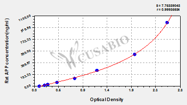
These standard curves are provided for demonstration only. A standard curve should be generated for each set of samples assayed.
ng/ml OD1 OD2 Average Corrected  
1000 2.557 2.520 2.539 2.429  
500 1.961 1.876 1.919 1.809  
250 1.197 1.204 1.201 1.091  
125 0.748 0.809 0.779 0.669  
62.5 0.436 0.446 0.441 0.331  
31.2 0.278 0.259 0.269 0.159  
15.6 0.206 0.209 0.208 0.098  
0 0.112 0.107 0.110    
Troubleshooting
and FAQs
Storage
Store at 2-8°C. Please refer to protocol.
Lead Time
3-5 working days after you place the order, and it takes another 3-5 days for delivery via DHL or FedEx
Description

This Rat APP ELISA Kit was designed for the quantitative measurement of Rat APP protein in serum, plasma, tissue homogenates . It is a Sandwich ELISA kit, its detection range is 15.6 ng/mL-1000 ng/mL and the sensitivity is 3.9 ng/mL.

Customer Reviews and Q&A

 Customer Reviews

There are currently no reviews for this product.

Submit a Review here

Target Background

Function
(From Uniprot)
Functions as a cell surface receptor and performs physiological functions on the surface of neurons relevant to neurite growth, neuronal adhesion and axonogenesis. Interaction between APP molecules on neighboring cells promotes synaptogenesis. Involved in cell mobility and transcription regulation through protein-protein interactions. Can promote transcription activation through binding to APBB1-KAT5 and inhibit Notch signaling through interaction with Numb. Couples to apoptosis-inducing pathways such as those mediated by G(O) and JIP. Inhibits G(o) alpha ATPase activity. Acts as a kinesin I membrane receptor, mediating the axonal transport of beta-secretase and presenilin 1. By acting as a kinesin I membrane receptor, plays a role in axonal anterograde transport of cargo towards synapes in axons. May be involved in copper homeostasis/oxidative stress through copper ion reduction. Can regulate neurite outgrowth through binding to components of the extracellular matrix such as heparin and collagen I and IV. The splice isoforms that contain the BPTI domain possess protease inhibitor activity. Induces a AGER-dependent pathway that involves activation of p38 MAPK, resulting in internalization of amyloid-beta peptide and leading to mitochondrial dysfunction in cultured mitochondrial dysfunction in cultured cortical neurons. Provides Cu(2+) ions for GPC1 which are required for release of nitric oxide (NO) and subsequent degradation of the heparan sulfate chains on GPC1.; Amyloid-beta peptides are lipophilic metal chelators with metal-reducing activity. Binds transient metals such as copper, zinc and iron. Rat and mouse amyloid-beta peptides bind only weakly transient metals and have little reducing activity due to substitutions of transient metal chelating residues. Amyloid-beta protein 42 may activate mononuclear phagocytes in the brain and elicits inflammatory responses. Promotes both tau aggregation and TPK II-mediated phosphorylation. Also binds GPC1 in lipid rafts.; Appicans elicit adhesion of neural cells to the extracellular matrix and may regulate neurite outgrowth in the brain.; The gamma-CTF peptides as well as the caspase-cleaved peptides, including C31, are potent enhancers of neuronal apoptosis.; N-APP binds TNFRSF21 triggering caspase activation and degeneration of both neuronal cell bodies (via caspase-3) and axons (via caspase-6).
Gene References into Functions
  1. These results, in Wistar rats, provide experimental support for the hypothesis that certain risk factors, such as energy metabolism dysfunction or the aging process itself, may increase vulnerability to beta-amyloid protein (Abeta). Hippocampal region is more susceptible to Abeta and its effect increases with age in relation to the neocortex. PMID: 29624358
  2. Abeta 25-35 was found to induce PC12 cell apoptosis in a dose-dependent manner (P<0.05). Moreover, Abeta 25-35 also caused dose-dependent disintegration of the cytoskeleton (P<0.05). PMID: 29436599
  3. Andrographolide (ANDRO) reduced inflammation-mediated neuronal damage by blocking inflammatory responses of microglial cells to Abeta(1-42), suggesting ANDRO may be an effective agent in modulating neuroinflammatory process in Alzheimer's disease. PMID: 28669260
  4. alanine in APP- intracellular domain(ICD) and the proline in APLP2-ICD lie directly behind a conserved caspase cleavage site PMID: 26921470
  5. These data suggest that methylation change in BDNF exon is involved in the regulation of BDNF expression by Abeta or SAM, and further support the view of specific epigenetic modifications of a certain BDNF gene transcript. PMID: 29305263
  6. Results from the present study indicated that pioglitazone may improve insulin sensitivity and ameliorate Abeta42 accumulation in rats with dietinduced IR by regulating AKT/GSK3beta activation, suggesting that pioglitazone may be a promising drug for AD treatment. PMID: 28447730
  7. Dynamic transport of Abeta aggregates is only observed between axon terminal and cell body. In addition to differential cellular uptake, more Abeta-peptide secretion is detected significantly from axons than from dendritic side. PMID: 28121396
  8. Results show that concerted signaling by both APP and Abeta is necessary for aberrant neuronal cell cycle entry. Thus, APP plays a central role in promotion of neurodegeneration, through activation of Ras-ERK signaling axis as well as GSK-3, and interfering with these signaling events would impede cell cycle reentry and neurodegeneration observed in Alzheimer's disease. PMID: 28374012
  9. Lynx1 and Abeta1-42 bind competitively to multiple nicotinic acetylcholine receptor subtypes. PMID: 27460145
  10. increased APP and/or beta-CTF impact the endocytic pathway to disrupt NGF trafficking and signaling, resulting in trophic deficits in basal forebrain cholinergic neurons. PMID: 27064279
  11. results establish a novel toxic impact ofAbeta oligomers on neuronal metabolism and suggest that Abeta oligomers-induced, NMDA receptor-mediated AMPK inhibition may play a key role in early brain metabolic defects in AD. PMID: 28302722
  12. The protein level of Abeta in serum and cerebrospinal fluid (CSF) was severity and timedependent during the acute phase in rats with traumatic spinal cord injury (SCI). Monitoring the level of Abeta protein in serum may improve the evaluation of SCI severity and the neuron functional status following SCI. PMID: 28259979
  13. O3 exposure (0.25 ppm) induced a significant increase of Syntaxin 5 and accumulation of betaA42 peptide in the reticulum in cells of the dentate gyrus PMID: 27366738
  14. results indicate that specifically "trapping" low-n oligomers provides a novel strategy for toxic Abeta42-oligomer recognition and removal. PMID: 26510576
  15. Results indicate that improving synaptic soluble AbetaPPalpha availability at synapses helps in reducing the functional NMDAR-related deregulation of hippocampal networks linked to aging PMID: 26402095
  16. High glucose leads to the increased expression of genes related to Abeta production, resulting in the accumulation of Abeta in the lens. PMID: 27028062
  17. Autoimmune Optic Neuritis Is Associated with Altered APP Cleavage in Neurons and Up-Regulation of p53 PMID: 26426258
  18. Redox-mediated posttranslational modification of brain proteins link Abeta and hyperglycaemia to cognitive dysfunction in metabolic syndrome/type 2 diabetes and Alzheimer disease. PMID: 26743041
  19. This study indicates that interfering intracellular Abeta especially mitochondrial Abeta accumulation, together with ameliorating Abeta-associated mitochondrial dysfunction, may contribute to the protective effects of huperzine A against Abeta neurotoxicity. PMID: 26024517
  20. Syt-1 and Syt-9 regulate endogenous APP-CTF and Abeta levels in PC12 cells. Secreted sAPPbeta levels were significantly reduced in PC12 cells lacking Syt-1 expression. PMID: 26202512
  21. Results show that downregulation of let-7d might contribute to isoflurane-induced learning and memory impairment through upregulating its target APP, and increasing the production of Abeta subsequently. PMID: 25799420
  22. Exposure to As-, Cd-, and Pb-mixture induces Abeta, amyloidogenic APP processing and cognitive impairments via oxidative stress-dependent neuroinflammation in young rats. PMID: 25288670
  23. Data suggests that APP is important in regulating intestinal cholesterol uptake in a fashion dependent upon specific proteolytic pathways. PMID: 25742317
  24. The soluble extracellular fragment of neuroligin-1 protects synapses in neurons damaged by ABETA oligomers. PMID: 26325471
  25. The present study indicates that Abeta-mediated Zn2+ influx into dentate granule cells, which may occur without AMPA receptor activation, transiently induces a short-term cognitive deficit. PMID: 25536033
  26. Incubation of brain homogenates with 70 microM hydrogen peroxide significantly influenced the profile of Abeta binding proteins and 0.1 mM isatin decreased the number of identified Abeta binding proteins. PMID: 25551598
  27. Abeta-40 Y10F exhibited remarkably decreased neurotoxicity compared to Abeta-40 which could be partly due to the reduced generation of hydrogen peroxide. PMID: 22754299
  28. The results of this study provide the first direct demonstration that AbetaPP is mostly distributed among glutamatergic rather than GABAergic or cholinergic terminals of the adult rat hippocampus. PMID: 24531160
  29. report we describe a novel neuroprotective function of mGlu3 receptors related to their ability to promote the non-amyloidogenic pathway of APP cleavage in astrocytes, thus enhancing sAPPalpha production PMID: 24291464
  30. Rodent-Abeta staining is present and increased after juvenile traumatic brain injury around cerebral blood microvessels, and the diameter of those is decreased by 25% and 34% at 2 and 6 months, respectively, without significant angiogenesis. PMID: 25052558
  31. Oxidative stress served as the key trigger to down-regulate miR-107 by cell- derived soluble Abeta. PMID: 24595404
  32. present results suggest that the amyloid peptide exhibits a protective antioxidant role in biological systems, but also has toxic actions independent of oxidative stress PMID: 23041348
  33. Both voltage-activated Ca2+ channel and NMDAR-dependent long-term potentiation in vivo are strongly inhibited by Abeta. PMID: 24298149
  34. the existence of a structurally related family of quasi-fibrillar conformers of Abeta42, which is stabilized both by curcumin and by Zn(2+.) PMID: 24599958
  35. analysis of the role of JIP1 in APP transport; knockdown of JIP1 did not affect either amyloid precursor protein transport or amyloid-beta peptide production PMID: 23825109
  36. Data suggest that the brain expression of neprilysin may promote amyloid beta-protein (Abeta)deposition in patients with Alzheimer's disease (AD). PMID: 24508800
  37. Neurodegeneration may occur in a retrograde fashion from axon terminals near beta-amyloid plaques which may spread through brain regions in cortical neuron cultures. PMID: 24114864
  38. APP is an independently operating cell adhesion molecule PMID: 23691241
  39. These data present novel insights into the subsynaptic localization of APPs PMID: 23815291
  40. Abeta42 globulomers have an antiparallel beta-sheet arrangement PMID: 23687299
  41. processing of amyloid precursor protein in cells undergoing apoptosis PMID: 23469123
  42. Identification of the minimal zinc-binding center in natural isoforms of amyloid-beta domain 1-16 using ESI-MS PMID: 23888782
  43. PuF may regulate the APP gene promoter PMID: 23368879
  44. results showed that EMP exposure can cause long-term impairment in impaired cognition and memory of rats, resulting in AD-like symptoms; this may be induced by enhancing oxidative stress and is related to autophagy dysfunction PMID: 23523687
  45. The CentA1-Ras-Elk-1 signaling pathway acts on mitochondria to regulate dendritic spine density and synaptic plasticity in response to Abeta in hippocampal neurons. PMID: 23516302
  46. Prion protein-mediated toxicity of amyloid-beta oligomers requires lipid rafts and the transmembrane LRP1 PMID: 23386614
  47. Neuronal binding and hippocampal neurotransmission were found to be suitable to account for the synaptotoxicity to different App assemblies, based on the stability of the applied App aggregates in these settings. PMID: 23374097
  48. Early brain injury alters the blood-brain barrier phenotype in parallel with beta-amyloid and cognitive changes in adulthood. PMID: 23149553
  49. S-nitrosylation pathway wherein Cdk5/NOS1 interaction enhances S-nitrosylation of Cdk5, mediates mitochondrial dysfunction and synaptic loss during the App etiology of Alzheimer's disease. PMID: 22874667
  50. C1q-induced LRP1B and GPR6 proteins expressed early in Alzheimer disease mouse models, are essential for the C1q-mediated protection against amyloid-beta neurotoxicity PMID: 23150673

Show More

Hide All

Subcellular Location
Cell membrane; Single-pass type I membrane protein. Membrane; Single-pass type I membrane protein. Perikaryon. Cell projection, growth cone. Membrane, clathrin-coated pit. Early endosome. Cytoplasmic vesicle.; [C83]: Endoplasmic reticulum. Golgi apparatus. Early endosome.; [C99]: Early endosome.; [Amyloid-beta protein 42]: Cell surface.; [Gamma-secretase C-terminal fragment 59]: Nucleus. Cytoplasm.; [Soluble APP-beta]: Secreted.
Protein Families
APP family
Tissue Specificity
Expressed in the brain. In the brain, non-L-APP isoforms are expressed in neurons, isoform APP695 being the predominant form. In astrocytes and microglial cells, almost 50% is L-isoform (appican).
Database Links
CUSABIO guaranteed quality
icon of phone
Call us
301-363-4651 (Available 9 a.m. to 5 p.m. CST from Monday to Friday)
icon of address
Address
7505 Fannin St., Ste 610, Room 7 (CUBIO Innovation Center), Houston, TX 77054, USA
icon of social media
Join us with

Subscribe newsletter

Leave a message

* To protect against spam, please pass the CAPTCHA test below.
CAPTCHA verification
© 2007-2024 CUSABIO TECHNOLOGY LLC All rights reserved. 鄂ICP备15011166号-1