Call us
301-363-4651 (Available 9 a.m. to 5 p.m. CST from Monday to Friday)
| Code | CSB-EP321366HIT |
| Abbreviation | Recombinant Human coronavirus 229E S protein, partial |
| MSDS | |
| Size | $224 |
| Order now | |
| Image |
|
| Have Questions? | Leave a Message or Start an on-line Chat |
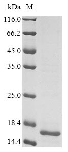
Recombinant Human coronavirus 229E Spike glycoprotein (S) is produced in E. coli and covers amino acids 785 to 873. The protein includes an N-terminal 10xHis-tag and a C-terminal Myc-tag, which makes purification and detection more straightforward. SDS-PAGE analysis confirms the purity level exceeds 85%, which should provide reliable results for research work.
The Spike glycoprotein (S) of Human coronavirus 229E appears to play a critical role in how the virus enters cells - it seems to handle both attachment to host cell receptors and the membrane fusion process. This protein has become a key target for researchers looking into viral infection mechanisms and possible therapeutic approaches. Understanding this protein may be crucial for grasping how coronaviruses interact with their hosts and for developing strategies against coronavirus-related diseases.
Potential Applications
Note: The applications listed below are based on what we know about this protein's biological functions, published research, and experience from experts in the field. However, we haven't fully tested all of these applications ourselves yet. We'd recommend running some preliminary tests first to make sure they work for your specific research goals.
The protein is expressed in E. coli as a partial Spike (S) fragment (785–873 aa) with dual tags (10xHis-N-terminal, Myc-C-terminal). While the His tag improves solubility, E. coli often struggles to produce correctly folded eukaryotic membrane-associated proteins like the S protein—even for fragments. No data confirms native secondary/tertiary structure (e.g., circular dichroism, thermal shift assays). E. coli may misfold the fragment or fail to form critical disulfide bonds (if present in 785–873 aa), leading to non-functional conformations. The fragment lacks the full-length S context (e.g., receptor-binding domain [RBD], fusion machinery), so it cannot recapitulate native interactions (e.g., with ACE2 or other S domains). Bioactivity (e.g., ligand binding, oligomerization) is untested and unlikely to mirror wild-type S.
1. Antibody Development and Epitope Mapping
This recombinant S fragment (785–873 aa) can serve as an immunogen for antibodies targeting this region, but antibody specificity must be validated against native S—E. coli-expressed protein may present non-native epitopes, leading to cross-reactivity. The His/Myc tags simplify purification/detection, but antibodies generated may not recognize the fragment in its native context (e.g., on viral particles or infected cells).
2. Protein-Protein Interaction Studies
Pull-down assays using the His tag can identify interactors, but results are highly dependent on correct folding—E. coli-expressed fragments often misfold, causing false positives/negatives. Identified partners must be validated via co-IP or functional assays to rule out artifacts. The fragment’s limited length (785–873 aa) restricts insights to interactions within this domain, not full-length S biology.
3. ELISA-Based Binding Assays
The dual-tagged protein can be used in ELISA to screen ligand/receptor interactions, but orientation and folding introduce variability—the His tag may force non-native immobilization, and misfolding could abolish binding. Results are qualitative at best; quantitative affinity measurements require confirmed native structure.
4. Structural and Biochemical Characterization
This fragment supports preliminary biophysical studies (e.g., CD for secondary structure, DLS for stability) but cannot inform native S architecture—E. coli-expressed protein may lack correct disulfide bonds or domain folding. Structural conclusions (e.g., oligomerization) must be contextualized by folding limitations.
Final Recommendation & Action Plan
This E. coli-expressed HCoV-229E S fragment (785–873 aa) has limited utility without rigorous validation first, confirming folding via CD spectroscopy and thermal shift assays to rule out misfolding; second, testing bioactivity (e.g., binding to known partners) to ensure native-like function. Optimize expression (e.g., co-express chaperones, lower induction temperature) to improve solubility. For antibody development, validate specificity against native S; for interactions/ELISA, use tag cleavage or orthogonal methods (e.g., SPR) to reduce artifacts. If folding/bioactivity fails, use a eukaryotic system (e.g., insect cells) to ensure native structure—this fragment alone is insufficient for reliable downstream applications without validation.
There are currently no reviews for this product.
KEGG: vg:918758

