Recombinant Severe acute respiratory syndrome coronavirus Spike glycoprotein (S), partial (Active)

In Stock
Code CSB-MP348663HQE
Abbreviation Recombinant SARS-CoV S protein, partial (Active)
MSDS
Size $138
Order now
Image
  • (Tris-Glycine gel) Discontinuous SDS-PAGE (reduced) with 5% enrichment gel and 15% separation gel.
  • Activity
    Measured by its binding ability in a functional ELISA. Immobilized SARS-CoV S-RBD at 2 μg/ml can bind Paguma larvata ACE2 (CSB-MP684964PAL), the EC50 is 5.056-7.559 ng/ml. Biological Activity Assay
  • Activity
    Measured by its binding ability in a functional ELISA. Immobilized SARS-CoV S-RBD at 5 μg/ml can bind human ACE2 (CSB-MP866317HU), the EC50 is 7.941-10.49 ng/ml. Biological Activity Assay
Have Questions? Leave a Message or Start an on-line Chat

Product Details

Purity
Greater than 95% as determined by SDS-PAGE.
Endotoxin
Less than 1.0 EU/ug as determined by LAL method.
Activity
①Measured by its binding ability in a functional ELISA. Immobilized SARS-CoV S-RBD at 2 μg/ml can bind Paguma larvata ACE2 (CSB-MP684964PAL), the EC50 is 5.056-7.559 ng/ml.②Measured by its binding ability in a functional ELISA. Immobilized SARS-CoV S-RBD at 5 μg/ml can bind human ACE2 (CSB-MP866317HU), the EC50 is 7.941-10.49 ng/ml.
Target Names
Uniprot No.
Research Area
Microbiology
Alternative Names
(S glycoprotein)(E2)(Peplomer protein)(Spike protein S1)(Spike protein S2)
Molecular Characterization
Species
Human SARS coronavirus (SARS-CoV) (Severe acute respiratory syndrome coronavirus)
Source
Mammalian cell
Expression Region
306-527aa
Target Protein Sequence
RVVPSGDVVRFPNITNLCPFGEVFNATKFPSVYAWERKKISNCVADYSVLYNSTFFSTFKCYGVSATKLNDLCFSNVYADSFVVKGDDVRQIAPGQTGVIADYNYKLPDDFMGCVLAWNTRNIDATSTGNYNYKYRYLRHGKLRPFERDISNVPFSPDGKPCTPPALNCYWPLNDYGFYTTTGIGYQPYRVVVLSFELLNAPATVCGPKLSTDLIKNQCVNF
Mol. Weight
30.0 kDa
Protein Length
Partial
Tag Info
N-terminal 10xHis-tagged and C-terminal Myc-tagged
Form
Lyophilized powder
Note: We will preferentially ship the format that we have in stock, however, if you have any special requirement for the format, please remark your requirement when placing the order, we will prepare according to your demand.
Buffer
Lyophilized from a 0.2 μm filtered 20 mM Tris-HCl, 0.5 M NaCl, 6% Trehalose, pH 8.0
Reconstitution
We recommend that this vial be briefly centrifuged prior to opening to bring the contents to the bottom. Please reconstitute protein in deionized sterile water to a concentration of 0.1-1.0 mg/mL.We recommend to add 5-50% of glycerol (final concentration) and aliquot for long-term storage at -20°C/-80°C. Our default final concentration of glycerol is 50%. Customers could use it as reference.
Troubleshooting and FAQs
Storage Condition
Store at -20°C/-80°C upon receipt, aliquoting is necessary for mutiple use. Avoid repeated freeze-thaw cycles.
Shelf Life
The shelf life is related to many factors, storage state, buffer ingredients, storage temperature and the stability of the protein itself.
Generally, the shelf life of liquid form is 6 months at -20°C/-80°C. The shelf life of lyophilized form is 12 months at -20°C/-80°C.
Lead Time
3-7 business days
Notes
Repeated freezing and thawing is not recommended. Store working aliquots at 4°C for up to one week.
Datasheet & COA
Please contact us to get it.
Description

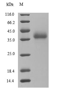
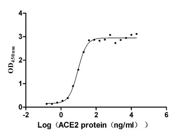
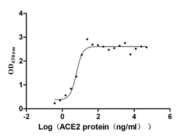
This recombinant protein corresponds to the receptor-binding domain (RBD, amino acids 306-527) of the SARS-CoV spike glycoprotein (S), expressed in mammalian cells with dual tags: an N-terminal 10×His tag and a C-terminal Myc tag for enhanced detection and purification. It demonstrates high purity (>95% by SDS-PAGE) and low endotoxin levels (<1.0 EU/μg, LAL method), ensuring suitability for sensitive microbiological assays. Functional validation via ELISA confirms its binding activity to both Paguma larvata ACE2 (CSB-MP684964PAL) and human ACE2 (CSB-MP866317HU), with EC50 of 5.056–7.559 ng/mL and 7.941–10.49 ng/mL, respectively, highlighting its utility in studying cross-species viral entry mechanisms and host receptor interactions. Provided as a lyophilized powder, this recombinant SARS-CoV S protein retains structural integrity and is optimized for reconstitution in downstream applications. Its mammalian expression ensures proper glycosylation and folding, making it a critical tool for SARS-CoV research, vaccine development, and therapeutic antibody screening in microbiology.

The spike (S) protein of SARS-CoV, a member of the coronavirus family, is a critical determinant of the virus's ability to infect host cells. This glycoprotein is integral to the virus's life cycle as it facilitates the initial attachment of the virus to host cells via binding to the ACE2 receptor. The S protein consists of two main subunits: the S1 subunit and the S2 subunit. The S1 subunit is primarily responsible for receptor recognition, while the S2 subunit plays a pivotal role in membrane fusion and the entry of the viral particle into the host cell [1][2].

The process of viral entry is dependent on several biochemical steps that include proteolytic cleavage of the S protein, which is essential for its fusion capability. In particular, cleavage at the S1/S2 site by host proteases such as the TMPRSS2 is crucial. This cleavage event activates the S protein, allowing it to facilitate membrane fusion between the viral and host cell membranes. Alternatively, in some cellular environments, other proteases such as Cathepsin B/L can also assist in this activation step [3][4][5]. Such proteolytic processing not only primes the S protein but also enhances the infectivity of the virus, underscoring the role of host factors in SARS-CoV and its related strains, including SARS-CoV-2 [4][6]. Moreover, the S protein is not only critical for the entry of the virus but also serves as a major target for vaccine development due to its immunogenic properties [1][2][7].

References:
[1] F. Cito, L. Amato, et al. A covid-19 hotspot area: activities and epidemiological findings, Microorganisms. vol. 8, no. 11, p. 1711, 2020. https://doi.org/10.3390/microorganisms8111711
[2] M. Medina-Enríquez, S. López‐León, J. Carlos-Escalante, Z. Aponte‐Torres, A. Cuapio, & T. Wegman‐Ostrosky. Ace2: the molecular doorway to sars-cov-2. Cell & Bioscience, vol. 10, no. 1, 2020. https://doi.org/10.1186/s13578-020-00519-8
[3] M. Hoffmann, H. Kleine‐Weber, & S. Pöhlmann. A multibasic cleavage site in the spike protein of sars-cov-2 is essential for infection of human lung cells. Molecular Cell, vol. 78, no. 4, p. 779-784.e5, 2020. https://doi.org/10.1016/j.molcel.2020.04.022
[4] M. Hoffmann, H. Kleine‐Weber, et al. Sars-cov-2 cell entry depends on ace2 and tmprss2 and is blocked by a clinically proven protease inhibitor. Cell, vol. 181, no. 2, p. 271-280.e8, 2020. https://doi.org/10.1016/j.cell.2020.02.052
[5] G. Sun, Y. Sui, et al. Structural basis of covalent inhibitory mechanism of tmprss2-related serine proteases by camostat. Journal of Virology, vol. 95, no. 19, 2021. https://doi.org/10.1128/jvi.00861-21
[6] Y. Park, J. Ahn, S. Hwang, K. Sung, J. Lim, & K. Kwack. Structural similarity analysis of the spike protein of sars-cov-2 and other sars-related coronaviruses. 2020. https://doi.org/10.20944/preprints202003.0409.v1
[7] S. Jangra, J. Vrieze, et al. Sterilizing immunity against sars-cov-2 infection in mice by a single-shot and modified imidazoquinoline tlr7/8 agonist-adjuvanted recombinant spike protein vaccine. 2020. https://doi.org/10.1101/2020.10.23.344085

Customer Reviews and Q&A

 Customer Reviews
Average Rating:
5.0 - 1 reviews

Submit a Review here

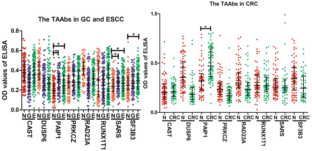
Applications : Recombinant proteins and the detection of TAAbs by ELISA

Review: All 15 proteins used in ELISA were purchased from CUSABIO (SF3B3, RUNX1T1, SARS, PAIP1, CAST, MAGEA12, CCDC6, RAD23A, NOL7, CRLF3, NAP1L4, SH2B1, LARP6, Wuhan, China). The scatter dot plot of the optical density (OD) values of ELISA for 8 TAAbs in the specific validation set.

By Anonymous

Target Background

Function
May down-regulate host tetherin (BST2) by lysosomal degradation, thereby counteracting its antiviral activity.; attaches the virion to the cell membrane by interacting with host receptor, initiating the infection. Binding to human ACE2 and CLEC4M/DC-SIGNR receptors and internalization of the virus into the endosomes of the host cell induces conformational changes in the S glycoprotein. Proteolysis by cathepsin CTSL may unmask the fusion peptide of S2 and activate membranes fusion within endosomes.; mediates fusion of the virion and cellular membranes by acting as a class I viral fusion protein. Under the current model, the protein has at least three conformational states: pre-fusion native state, pre-hairpin intermediate state, and post-fusion hairpin state. During viral and target cell membrane fusion, the coiled coil regions (heptad repeats) assume a trimer-of-hairpins structure, positioning the fusion peptide in close proximity to the C-terminal region of the ectodomain. The formation of this structure appears to drive apposition and subsequent fusion of viral and target cell membranes.; Acts as a viral fusion peptide which is unmasked following S2 cleavage occurring upon virus endocytosis.
Subcellular Location
Virion membrane; Single-pass type I membrane protein. Host endoplasmic reticulum-Golgi intermediate compartment membrane; Single-pass type I membrane protein. Host cell membrane; Single-pass type I membrane protein.
Protein Families
Betacoronaviruses spike protein family
CUSABIO guaranteed quality
icon of phone
Call us
301-363-4651 (Available 9 a.m. to 5 p.m. CST from Monday to Friday)
icon of address
Address
7505 Fannin St., Ste 610, Room 7 (CUBIO Innovation Center), Houston, TX 77054, USA
icon of social media
Join us with

Subscribe newsletter

Leave a message

* To protect against spam, please pass the CAPTCHA test below.
CAPTCHA verification
© 2007-2025 CUSABIO TECHNOLOGY LLC All rights reserved. 鄂ICP备15011166号-1
Place an order now

I. Product details

*
*
*
*

II. Contact details

*
*

III. Ship To

*
*
*
*
*
*
*

IV. Bill To

*
*
*
*
*
*
*
*