Call us
301-363-4651 (Available 9 a.m. to 5 p.m. CST from Monday to Friday)
| Code | CSB-YP3324GMY2 |
| Abbreviation | Recombinant SARS-CoV-2 S protein, partial |
| MSDS | |
| Size | $250 |
| Order now | |
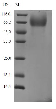
| Image |
|
| Have Questions? | Leave a Message or Start an on-line Chat |
The Recombinant Severe acute respiratory syndrome coronavirus 2 Spike glycoprotein (S) is expressed in a yeast system, covering the amino acid region 16-318. It features an N-terminal 6xHis-PDI tag for enhanced purification and detection. The protein is provided at a purity level greater than 90%, as confirmed by SDS-PAGE analysis, ensuring reliable results in research applications.
SARS-CoV-2's Spike glycoprotein (S) appears to be essential for viral entry into host cells, mediating attachment and fusion processes. It likely plays a critical role in the viral life cycle and has become a major target for vaccine development and therapeutic interventions. Understanding its structure and function may be crucial for advancing research on coronavirus pathogenesis and potential treatments.
Potential Applications
Note: The applications listed below are based on what we know about this protein's biological functions, published research, and experience from experts in the field. However, we haven't fully tested all of these applications ourselves yet. We'd recommend running some preliminary tests first to make sure they work for your specific research goals.
The protein is expressed in yeast, a eukaryotic system that supports better disulfide bond formation and solubility than E. coli, aided by the N-terminal PDI tag (protein disulfide isomerase, which catalyzes correct disulfide pairing). However, it is a partial fragment (16–318 aa) of the spike (S) protein, so folding reflects only this region—not the full-length S trimer. While PDI improves disulfide-dependent folding, there is no direct evidence of native conformation for the fragment (e.g., circular dichroism for secondary structure, thermal shift assays for stability, or reactivity with conformation-specific antibodies). Bioactivity is limited to the fragment: it may retain antigenicity for the 16–318 aa region (useful for antibody screening) but cannot mimic full-length S functions (e.g., ACE2 binding, which requires the receptor-binding domain, RBD, ~319–541 aa). The yeast system provides eukaryotic post-translational modifications (PTMs), but these differ from human cells, potentially affecting interactions.
1. Antibody Development and Screening
This recombinant SARS-CoV-2 spike fragment (16–318 aa) can serve as an antigen for generating antibodies targeting this specific region (not full-length S or the RBD). The N-terminal 6xHis-PDI tag simplifies purification and immobilization for immunization/screening. ELISA with this protein may identify antibodies against the 16–318 aa fragment, but results do not guarantee reactivity with full-length S or the RBD. High purity (>90%) supports consistent antigen presentation, but antibody specificity must be validated against the fragment (e.g., via Western blot or peptide mapping).
2. Protein-Protein Interaction Studies
Pull-down assays using the 6xHis-PDI tag can explore interactions with partners targeting the 16–318 aa region. Immobilization on nickel matrices and incubation with lysates/purified proteins may identify interactors, but yeast eukaryotic PTMs are not human-specific—interactions observed may not recapitulate native human cell biology. The partial fragment limits studies to interactions within this domain; full-length S or RBD interactions cannot be inferred.
3. Structural and Biochemical Characterization
This fragment is suitable for fragment-specific structural/biochemical studies (e.g., CD for secondary structure, DLS for stability, or thermal shift assays). High purity enables biophysical analyses, but results reflect only the 16–318 aa region—interpretation requires caution, as isolated fragments often misfold or behave differently than the full-length protein. X-ray crystallography/NMR/cryo-EM of the fragment may reveal local structure but not full-length S architecture.
4. ELISA-Based Binding Assays
Direct or capture ELISA formats can quantify binding of ligands/small molecules to the 16–318 aa fragment. Sandwich ELISA (capturing via anti-His antibodies) may preserve fragment orientation, but results are fragment-specific—binding to full-length S or the RBD cannot be extrapolated. The tag facilitates standardization, but assay validation (e.g., using fragment-specific antibodies) is critical to avoid misinterpreting full-length S activity.
Final Recommendation & Action Plan
This yeast-expressed spike fragment (16–318 aa) with a 6xHis-PDI tag has potential for fragment-specific applications (antibody generation, biochemical characterization) but requires rigorous validation: first, confirm folding of the 16–318 aa region using CD spectroscopy or thermal shift assays; second, validate antigenicity via ELISA with fragment-specific antibodies; third, acknowledge limitations (partial fragment, non-human PTMs) in downstream use. For interaction studies, pair with human cell-based assays to confirm physiological relevance. If folding/antigenicity are confirmed, use the protein for its intended fragment-specific goals—avoid overinterpreting results as representative of full-length S. If folding fails, optimize expression/purification (e.g., adjust PDI co-expression) or use a system with more native-like folding (e.g., mammalian cells for full-length S).
There are currently no reviews for this product.

