Recombinant Human respiratory syncytial virus A Fusion glycoprotein F0 (F), partial

In Stock
Code CSB-EP356041HPOa0
Abbreviation Recombinant Human respiratory syncytial virus A Fusion glycoprotein F0, partial
MSDS
Size $224
Order now
Image
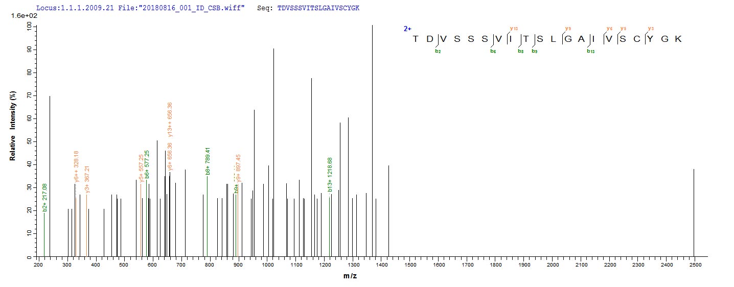
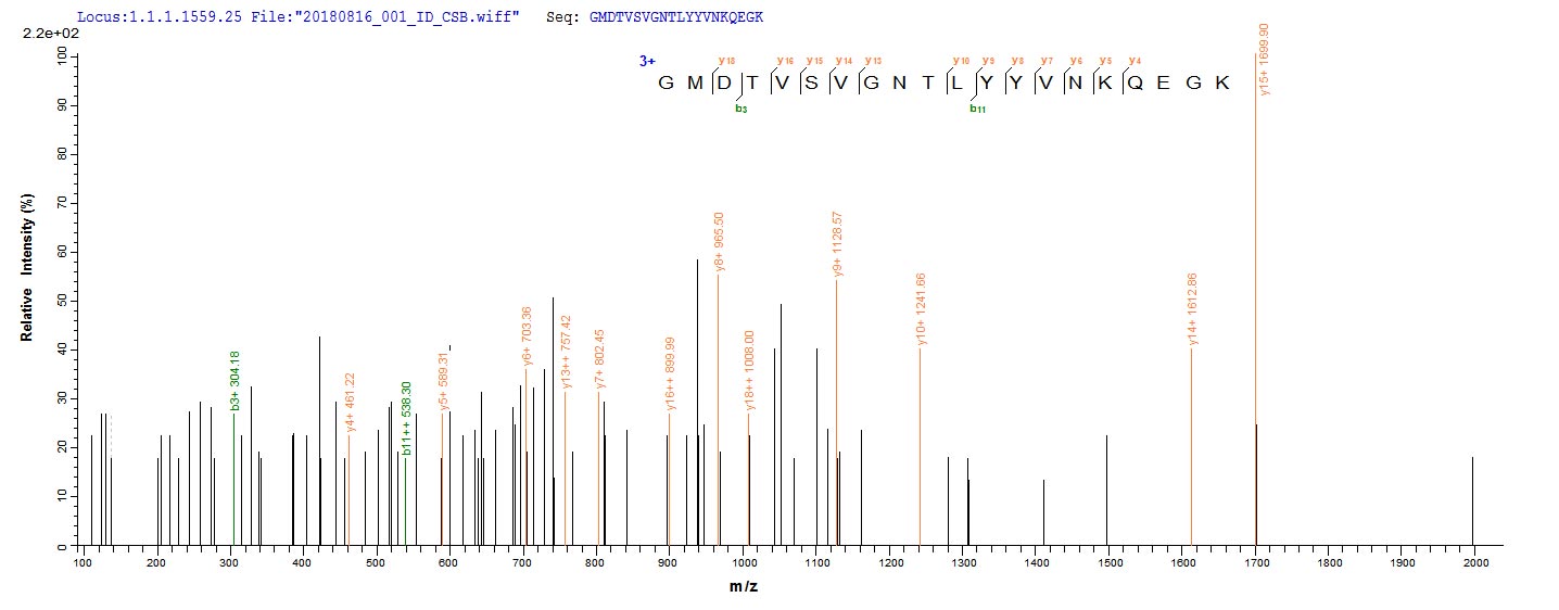
  • Based on the SEQUEST from database of E.coli host and target protein, the LC-MS/MS Analysis result of CSB-EP356041HPOa0 could indicate that this peptide derived from E.coli-expressed Human respiratory syncytial virus A (strain A2) F.
  • Based on the SEQUEST from database of E.coli host and target protein, the LC-MS/MS Analysis result of CSB-EP356041HPOa0 could indicate that this peptide derived from E.coli-expressed Human respiratory syncytial virus A (strain A2) F.
Have Questions? Leave a Message or Start an on-line Chat

Product Details

Purity
Greater than 85% as determined by SDS-PAGE.
Target Names
F
Uniprot No.
Research Area
Signal Transduction
Alternative Names
F; Fusion glycoprotein F0; Protein F) [Cleaved into: Fusion glycoprotein F2'; Interchain peptide; Fusion glycoprotein F2; Fusion glycoprotein F1]
Species
Human respiratory syncytial virus A (strain A2)
Source
E.coli
Expression Region
27-529aa
Target Protein Sequence
NITEEFYQSTCSAVSKGYLSALRTGWYTSVITIELSNIKENKCNGTDAKVKLIKQELDKYKNAVTELQLLMQSTPPTNNRARRELPRFMNYTLNNAKKTNVTLSKKRKRRFLGFLLGVGSAIASGVAVSKVLHLEGEVNKIKSALLSTNKAVVSLSNGVSVLTSKVLDLKNYIDKQLLPIVNKQSCSISNIETVIEFQQKNNRLLEITREFSVNAGVTTPVSTYMLTNSELLSLINDMPITNDQKKLMSNNVQIVRQQSYSIMSIIKEEVLAYVVQLPLYGVIDTPCWKLHTSPLCTTNTKEGSNICLTRTDRGWYCDNAGSVSFFPQAETCKVQSNRVFCDTMNSLTLPSEINLCNVDIFNPKYDCKIMTSKTDVSSSVITSLGAIVSCYGKTKCTASNKNRGIIKTFSNGCDYVSNKGMDTVSVGNTLYYVNKQEGKSLYVKGEPIINFYDPLVFPSDEFDASISQVNEKINQSLAFIRKSDELLHNVNAGKSTTNIMITT
Note: The complete sequence may include tag sequence, target protein sequence, linker sequence and extra sequence that is translated with the protein sequence for the purpose(s) of secretion, stability, solubility, etc.
If the exact amino acid sequence of this recombinant protein is critical to your application, please explicitly request the full and complete sequence of this protein before ordering.
Mol. Weight
58.9 kDa
Protein Length
Extracellular Domain
Tag Info
N-terminal 6xHis-tagged
Form
Liquid or Lyophilized powder
Note: We will preferentially ship the format that we have in stock, however, if you have any special requirement for the format, please remark your requirement when placing the order, we will prepare according to your demand.
Buffer
If the delivery form is liquid, the default storage buffer is Tris/PBS-based buffer, 5%-50% glycerol.
Note: If you have any special requirement for the glycerol content, please remark when you place the order.
If the delivery form is lyophilized powder, the buffer before lyophilization is Tris/PBS-based buffer, 6% Trehalose.
Reconstitution
We recommend that this vial be briefly centrifuged prior to opening to bring the contents to the bottom. Please reconstitute protein in deionized sterile water to a concentration of 0.1-1.0 mg/mL.We recommend to add 5-50% of glycerol (final concentration) and aliquot for long-term storage at -20°C/-80°C. Our default final concentration of glycerol is 50%. Customers could use it as reference.
Troubleshooting and FAQs
Storage Condition
Store at -20°C/-80°C upon receipt, aliquoting is necessary for mutiple use. Avoid repeated freeze-thaw cycles.
Shelf Life
The shelf life is related to many factors, storage state, buffer ingredients, storage temperature and the stability of the protein itself.
Generally, the shelf life of liquid form is 6 months at -20°C/-80°C. The shelf life of lyophilized form is 12 months at -20°C/-80°C.
Lead Time
3-7 business days
Notes
Repeated freezing and thawing is not recommended. Store working aliquots at 4°C for up to one week.
Datasheet & COA
Please contact us to get it.
Description

Recombinant Human respiratory syncytial virus A Fusion glycoprotein F0 is produced in E. coli and contains the extracellular domain from amino acids 27 to 529. The protein carries an N-terminal 6xHis-tag, which helps with purification and detection. SDS-PAGE analysis shows purity levels greater than 85%, making it appropriate for various research applications. Low endotoxin levels are maintained in the final product, which appears important for sensitive experimental work.

The Fusion glycoprotein F0 of Human respiratory syncytial virus A seems to play a central role in how the virus infects cells by mediating membrane fusion and viral entry into host cells. This protein has become a key target for research into viral pathogenesis and vaccine development, since it likely initiates the infection process. Understanding how this protein works may be critical for developing therapeutic interventions and grasping the mechanisms behind viral entry.

Potential Applications

Note: The applications listed below are based on what we know about this protein's biological functions, published research, and experience from experts in the field. However, we haven't fully tested all of these applications ourselves yet. We'd recommend running some preliminary tests first to make sure they work for your specific research goals.

Human respiratory syncytial virus A Fusion glycoprotein F0 is a complex viral glycoprotein that requires precise folding, proper glycosylation at multiple asparagine residues, correct disulfide bond formation, and proteolytic cleavage for its functional activity in membrane fusion and viral entry. The E. coli expression system cannot provide the eukaryotic folding environment, glycosylation machinery, or proteolytic processing required for this viral glycoprotein. The extracellular domain (27-529aa) lacks the transmembrane and cytoplasmic regions, but the absence of glycosylation and potential improper disulfide bonding means the protein is unlikely to adopt its native conformation. The N-terminal 6xHis-tag may cause minimal steric interference, but the probability of correct folding with functional activity is extremely low.

1. Antibody Development and Characterization Studies

This application has significant limitations. While antibodies can be generated against linear epitopes, the non-glycosylated, misfolded protein may not present conformational epitopes found on the native, glycosylated F protein on RSV virions. Antibodies raised against this recombinant protein may not efficiently neutralize the virus or recognize the native protein in immunological assays.

2. Structural and Biophysical Characterization

Basic biophysical analysis can be performed but will not reflect native F protein structure. Techniques like circular dichroism spectroscopy may assess secondary structure content, but the lack of glycosylation and potential improper disulfide bonding mean results will describe an artificial protein rather than the viral glycoprotein. The His-tag may interfere with high-resolution structural studies.

Final Recommendation & Action Plan

This E. coli-expressed RSV F protein extracellular domain is unsuitable for functional studies due to the essential requirements for glycosylation and proper folding that cannot be met in this expression system. The protein should not be used for interaction studies and vaccine research. Application 1 (antibody development) has severe limitations and may produce antibodies with poor viral neutralization capability. Application 2 provides only basic characterization of the misfolded protein. For reliable RSV research, use full-length, glycosylated F protein expressed in mammalian or insect cell systems that support proper post-translational modifications and preserve native conformational epitopes.

Customer Reviews and Q&A

 Customer Reviews

There are currently no reviews for this product.

Submit a Review here

Target Background

Function
Inactive precursor that is cleaved at two sites by a furin-like protease to give rise to the mature F1 and F2 fusion glycoproteins.; Class I viral fusion protein. Under the current model, the protein has at least 3 conformational states: pre-fusion native state, pre-hairpin intermediate state, and post-fusion hairpin state. During viral and plasma cell membrane fusion, the coiled coil regions assume a trimer-of-hairpins structure, positioning the fusion peptide in close proximity to the C-terminal region of the ectodomain. The formation of this structure appears to drive apposition and subsequent fusion of viral and cellular membranes leading to delivery of the nucleocapsid into the cytoplasm. This fusion is pH independent and occurs at the plasma or endosomal membrane (Probable). The trimer of F1-F2 (F protein) also facilitates the attachment to host cell by binding to host heparan sulfate. F protein is involved in the entry into the host cell through the interaction with host IGFR1. This interaction activates PRKCZ/PKCzeta that recruits host NCL/nucleolin to the apical cell surface where it can bind fusion glycoprotein F1. Later in infection, F protein expressed at the plasma membrane of infected cells can mediate fusion with adjacent cells to form syncytia, a cytopathic effect that could lead to tissue necrosis. F protein may trigger p53-dependent apoptosis.; Major determinant of the species specificity of RSV infection. The trimer of F1-F2 (F protein) also facilitates the attachment to host cell by binding to host heparan sulfate. F protein is involved in the entry into the host cell through the interaction with host IGFR1. This interaction activates PRKCZ/PKCzeta that recruits host NCL/nucleolin to the apical cell surface where it can bind fusion glycoprotein F1. Later in infection, F protein expressed at the plasma membrane of infected cells can mediate fusion with adjacent cells to form syncytia, a cytopathic effect that could lead to tissue necrosis. F protein seems to trigger p53-dependent apoptosis.
Subcellular Location
[Fusion glycoprotein F0]: Host Golgi apparatus membrane; Single-pass membrane protein.; [Fusion glycoprotein F1]: Virion membrane; Single-pass type I membrane protein. Host cell membrane; Single-pass membrane protein.; [Fusion glycoprotein F2]: Virion membrane. Host cell membrane.
Protein Families
Paramyxoviruses fusion glycoprotein family
CUSABIO guaranteed quality
icon of phone
Call us
301-363-4651 (Available 9 a.m. to 5 p.m. CST from Monday to Friday)
icon of address
Address
7505 Fannin St., Ste 610, Room 7 (CUBIO Innovation Center), Houston, TX 77054, USA
icon of social media
Join us with

Subscribe newsletter

Leave a message

* To protect against spam, please pass the CAPTCHA test below.
CAPTCHA verification
© 2007-2025 CUSABIO TECHNOLOGY LLC All rights reserved. 鄂ICP备15011166号-1
×
Place an order now

I. Product details

*
*
*
*

II. Contact details

*
*

III. Ship To

*
*
*
*
*
*
*

IV. Bill To

*
*
*
*
*
*
*
*