Call us
301-363-4651 (Available 9 a.m. to 5 p.m. CST from Monday to Friday)
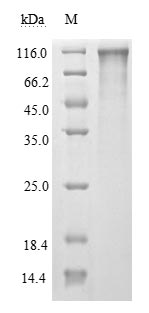
| Code | CSB-MP3324GMY |
| Abbreviation | Recombinant SARS-CoV-2 S protein, partial (Active) |
| MSDS | |
| Size | $256 |
| Order now | |
| Image |
|
| Have Questions? | Leave a Message or Start an on-line Chat |
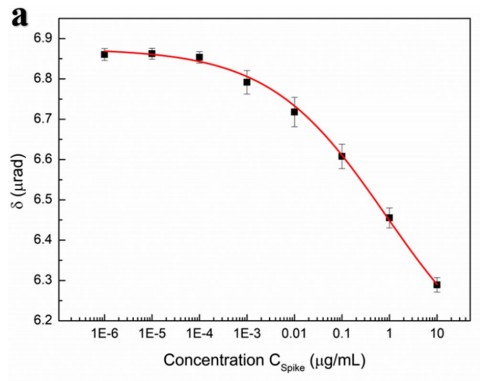
Applications : MO CM biochips
Review: Dependence of the concentration of spike glycoprotein Cspike (μg/ml) with respect to the CM rotation angle δ (μrad) form our MO CM biochips (the red line in the fgure is the ftting curve of the logistic function).
By Anonymous
Applications : Immunoblot analysis
Review: IL-1B release at 24 h following spike protein challenge in murine alveolar macrophages in the absence or presence of PPAR-α antagonist MK886 3µM. The results are expressed as mean ±SD of n = 4 experiments performed in triplicate. *** p < 0.001; ** p < 0.01, and * p < 0.05.
By Anonymous
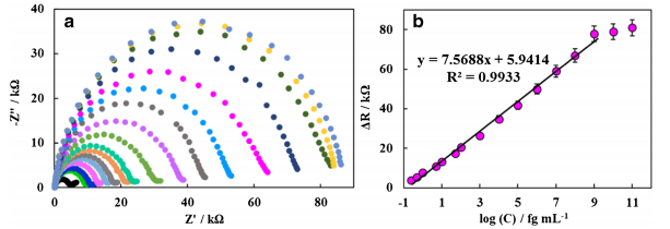
Applications : EIS responses of the designed biodevice
Review: EIS responses of the designed biodevice after incubation with different spike proteins with different concentrations.
By Anonymous
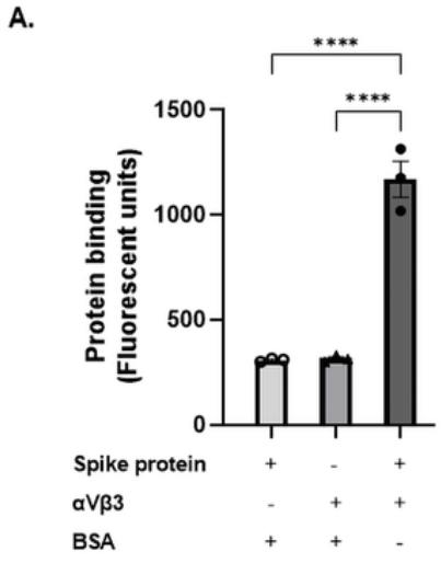
Applications : fluorescent-based protein assay
Review: Recombinant SARS-CoV-2 spike protein was immobilised and its binding to recombinant αVβ3 protein was measured using an anti-αVβ3 fluorescent antibody.
By Anonymous
Applications : /
Review: This project investigated the interactions of fatty acid derivatives and the SARS-CoV-2 spike fragment (FKNIDGYFKI decapeptide) on Aβ42 aggregation.
By Anonymous

